
Leukocyte Adhesion Cascade
Study all steps of the leukocyte adhesion cascade with one system
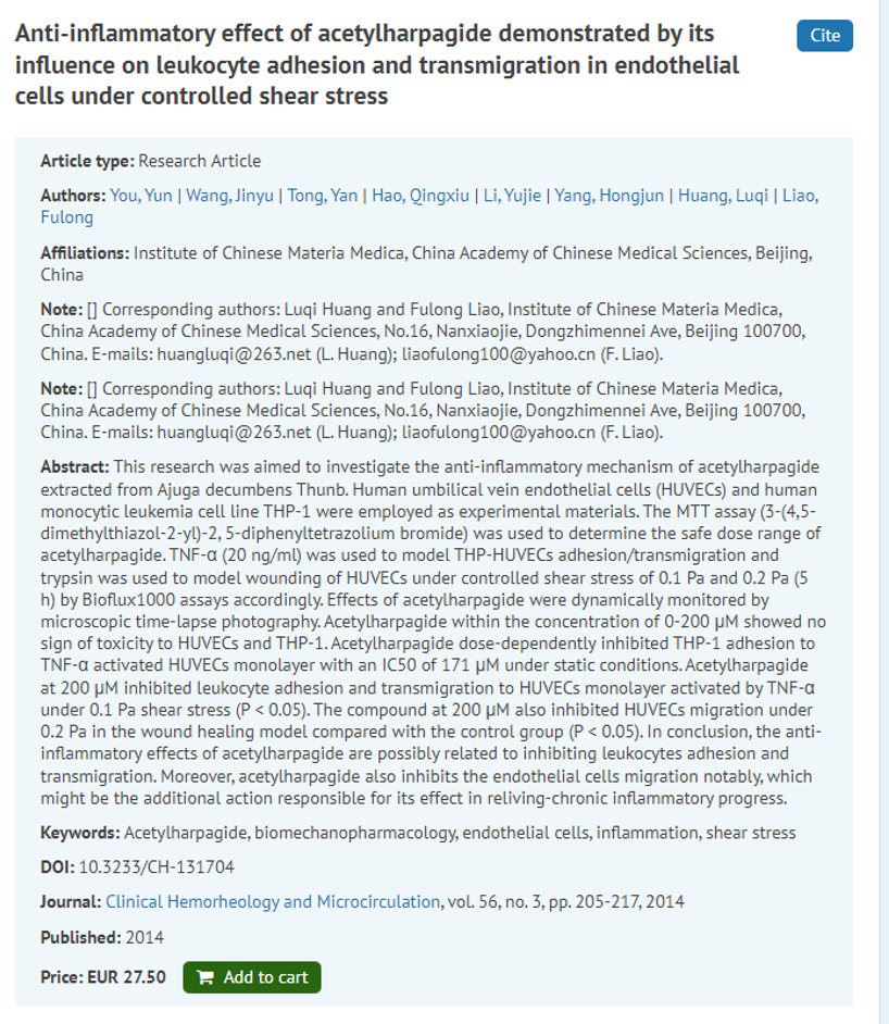
The leukocyte adhesion cascade consists of capture, rolling, slow rolling, arrest, strengthening/spreading, and transmigration. Typically, analyses of these steps require at least 3 separate systems and/or assays.
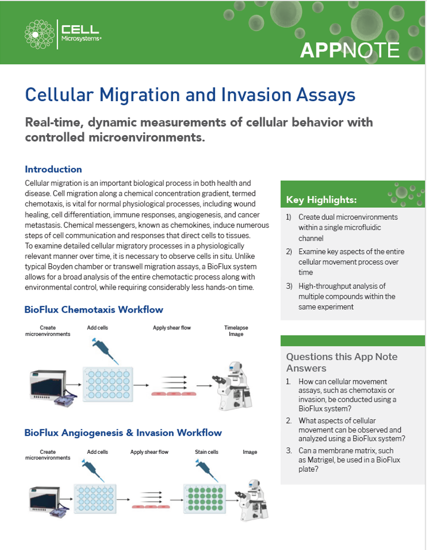
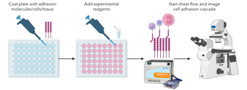
BioFlux shear flow systems enable the visualization and analysis of all of the steps of the leukocyte adhesion cascade under physiological/pathological shear flow using one system.
General BioFlux workflow for cell adhesion cascade assays
Key BioFlux Advantages For Cell Adhesion Cascade Studies
- Image and quantify live cell adhesion and rolling in a physiological environment using primary endothelial cells, human peripheral blood mononuclear cells, and more
- Utilize time-lapse microscopy and powerful post-experiment image analysis software to determine rolling distance under flow
- Examine up tp 24 different monolayers at one time to determine IC50 concentrations or screen multiple compounds
- Study truly biological cell adhesion and transmigration in the same assay
Leukocyte adhesion cascade assays using BioFlux
Adhesion
Rolling
Transmigration
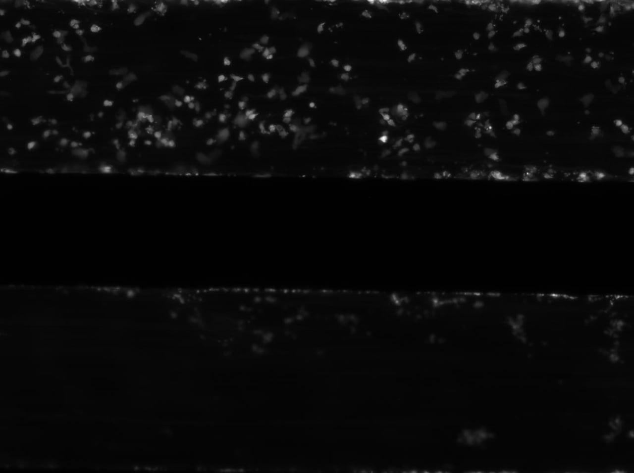
Recombinant human vimentin binds to P-selectin and blocks neutrophil capture and rolling on platelets and endothelium
The top channel shows the inhibition of neutrophil adhesion by recombinant human vimentin. The bottom channel shows neutrophil adhesion under control conditions.
The vimentin rod domain blocks P-selectin-P-selectin glycoprotein ligand 1 interactions to attenuate leukocyte adhesion to inflamed endothelium
Neutrophil rolling on endothelial cells under shear flow. Scale bar = 50 µm
Ezrin and Moesin Are Required for Efficient T Cell Adhesion and Homing to Lymphoid Organs
Tracking of the transmigration of CMTMR stained wild-type T cells (red) and CFSE stained ERM-deficient T cells (green) through TNF-α treated 3B-11 endothelial cells.Â