
Our Six Clone Challenge Recipients
Here, we spotlight the remarkable journeys of the six recipients of our Clone Challenge Grant Giveaway. Delve into their unique narratives to learn about the clone challenges these researchers faced. Then, discover how the innovative capabilities of CellRaftA microscale polystyrene growth surface within an array used to spatially segregate cells/colonies while maintaining shared media access, enabling imaging over time and targeted isolation. More Technology were used to successfully navigate and overcome these obstacles.
The Challenge
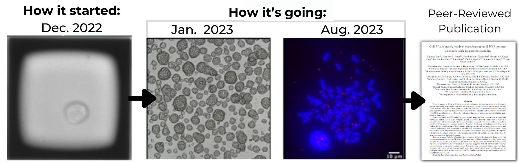
GBM39 is a human glioblastoma cell line, which is cultured in suspension as spheroids to prevent differentiation.
Extra-chromosomal DNAs (ecDNAs) frequently occur across many malignant cancer types and drive cancer evolution. GBM39 is good model cell line to study ecDNA as it has simple and high copy number of ecDNAs.
However, GBM39 is not homogeneous, and 90% of the cells (GBM39-ec) have EGFR amplicons on ecDNAs, while 10% of the cells (GBM39-hsr) have EGFR amplicons on homogeneously staining regions (HSRs). Additionally, as GBM39 grows slowly in suspension, it is challenging to grow many clones for screening.
The researcher wanted to isolate GBM39-ec and GBM39-hsr monoclonal populations from the heterogenous GBM39 cell line to screen using Fluorescence In Situ Hybridization (FISH) in metaphase spread and obtain isogenic cell lines that have EGFR on ecDNAs or HSRs.
A Success Story
GBM39 cells were seeded on a CellRaftA microscale polystyrene growth surface within an array used to spatially segregate cells/colonies while maintaining shared media access, enabling imaging over time and targeted isolation. More® Array, and 30 clones were isolated and expanded into ultra-low attachment plates to form spheroids.
In only one month, clones were able to be derived and were delivered back to the researcher. Upon receiving the colonies, Fluorescence In Situ Hybridization (FISH) in metaphase chromosome spread slides confirmed clones with both desired genotypes. Since then, a peer-reviewed publication is in progress and a preprint is available HERE.
The Challenge
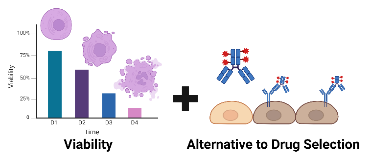
The researcher delivered polyclonal gene-edited populations of Edited 1 and Edited 2 suspension cell lines to Cell Microsystems to form monoclonal colonies of desired edits. These cells require neighboring populations to grow from single clones, and as a result, the researcher has mostly relied on drug-resistance markers to avoid this hurdle.
The researcher wanted an alternative cloning process without stressful drug selection.
A Success Story
Cells were seeded on a CellRaftA microscale polystyrene growth surface within an array used to spatially segregate cells/colonies while maintaining shared media access, enabling imaging over time and targeted isolation. More® Array and adhered to the surface using Poly-L-Lysine (PLL). Thousands of single cells were identified and screened using the CellRaftA microscale polystyrene growth surface within an array used to spatially segregate cells/colonies while maintaining shared media access, enabling imaging over time and targeted isolation. More® Cytometry software.
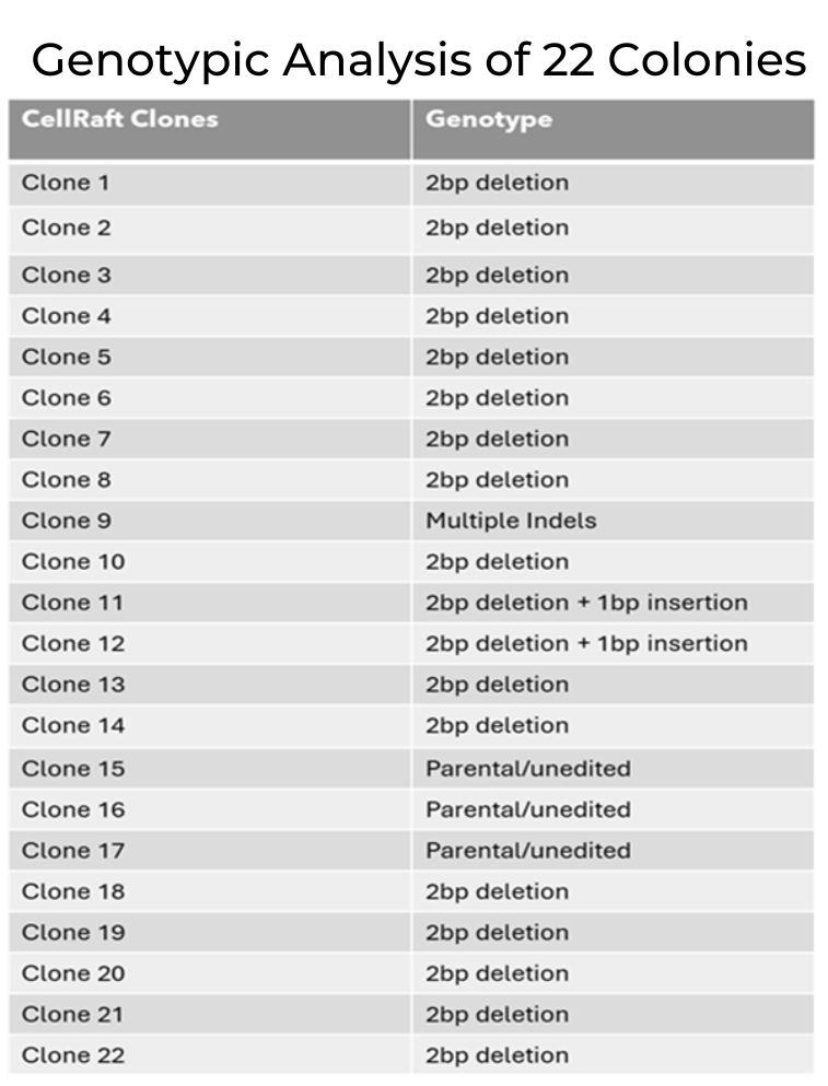
Of the clones identified, 22 clones were expanded and delivered back to the researcher to confirm monoclonalityEvidence that a colony/line originated from one cell; often supported by time-course imaging and traceability. More and that cells contained the edit of interest. All 22 sent were confirmed to be monoclonal, with 86% containing an indel of interest.
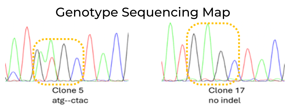
2bp deletion in Clone 5 compared to no indels in Clone 17.
All 22 colonies were monoclonal; 19 contained an indel of interest.
The Challenge
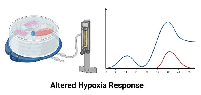
To study hypoxia-induced gene expression, researchers at an academic lab needed to generate monoclonal populations of Hep3B human hepatoma cells. However, when cells were plated in limiting dilutionA statistical cloning approach where cells are diluted to low density to “hopefully” seed one cell per well; commonly used but can be low-efficiency for difficult-to-clone lines. More, populations that arose from a small number of cells (1-3 cells) exhibited a permanently altered hypoxia response.
The researchers turned to the Cell Microsystems Clone Challenge to leverage CellRaftA microscale polystyrene growth surface within an array used to spatially segregate cells/colonies while maintaining shared media access, enabling imaging over time and targeted isolation. More Technology for high-efficiency single cell cloning that could return clonal populations of cells with their desired phenotype.
A Success Story
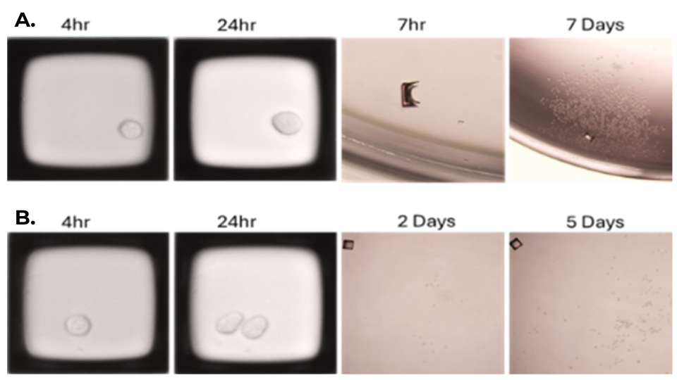
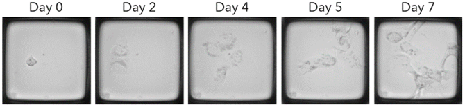
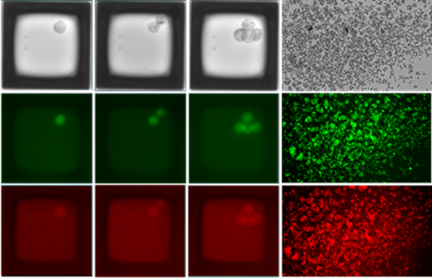
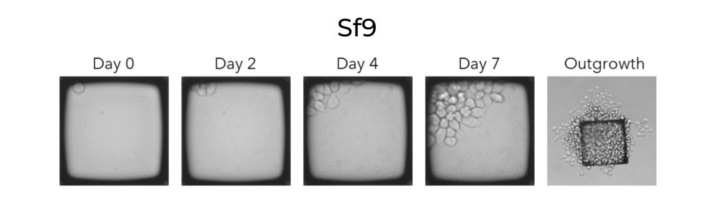
Representative time course images of Hep3B cells proliferating from a single cell to a monoclonal colony on a 200S CellRaftA microscale polystyrene growth surface within an array used to spatially segregate cells/colonies while maintaining shared media access, enabling imaging over time and targeted isolation. More® Arrays. CellRafts of interest were automatically isolated on the CellRaftA microscale polystyrene growth surface within an array used to spatially segregate cells/colonies while maintaining shared media access, enabling imaging over time and targeted isolation. More AIR® System from the 200S Array after 7 days.
Cells were seeded on an Array forming hundreds of clonal colonies. Colonies were screened using CellRaftA microscale polystyrene growth surface within an array used to spatially segregate cells/colonies while maintaining shared media access, enabling imaging over time and targeted isolation. More® Cytometry software and 68 clones were isolated. 18 of the clones were further expanded into flasks and delivered back to the researcher for analysis.
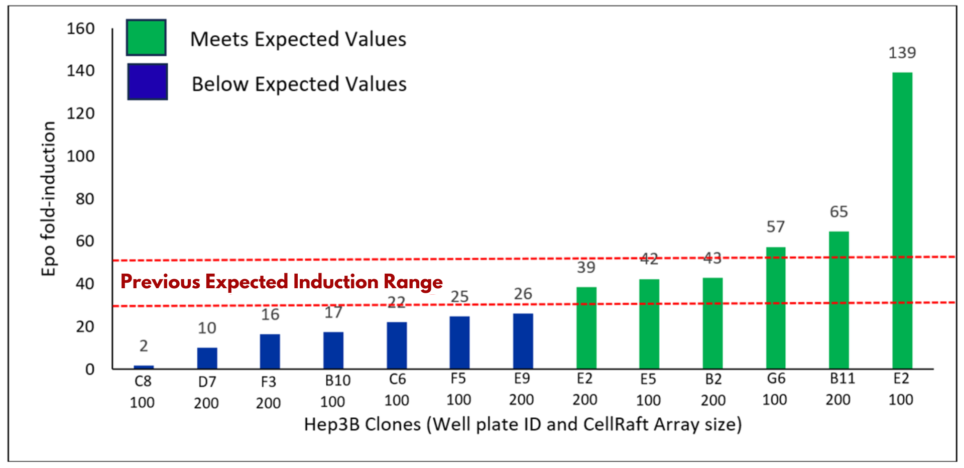
RT-PCR of hypoxia-induced clones revealed a >30-fold induction of Epo in 6 out of 13 clones tested, a phenotype that was previously lost when seeding cells at low densities in traditional single cell cloning workflows. The generation of clonal populations, from seeding the CellRaft ArrayConsumables containing thousands of microwells (containing CellRafts) for spatial segregation, imaging, and isolation of single cells, monoclonal colonies, or 3D cultures. More to expansion of clones in 96-well plates, required less than 3 weeks of total workflow time and less than 2 hours of hands-on time.
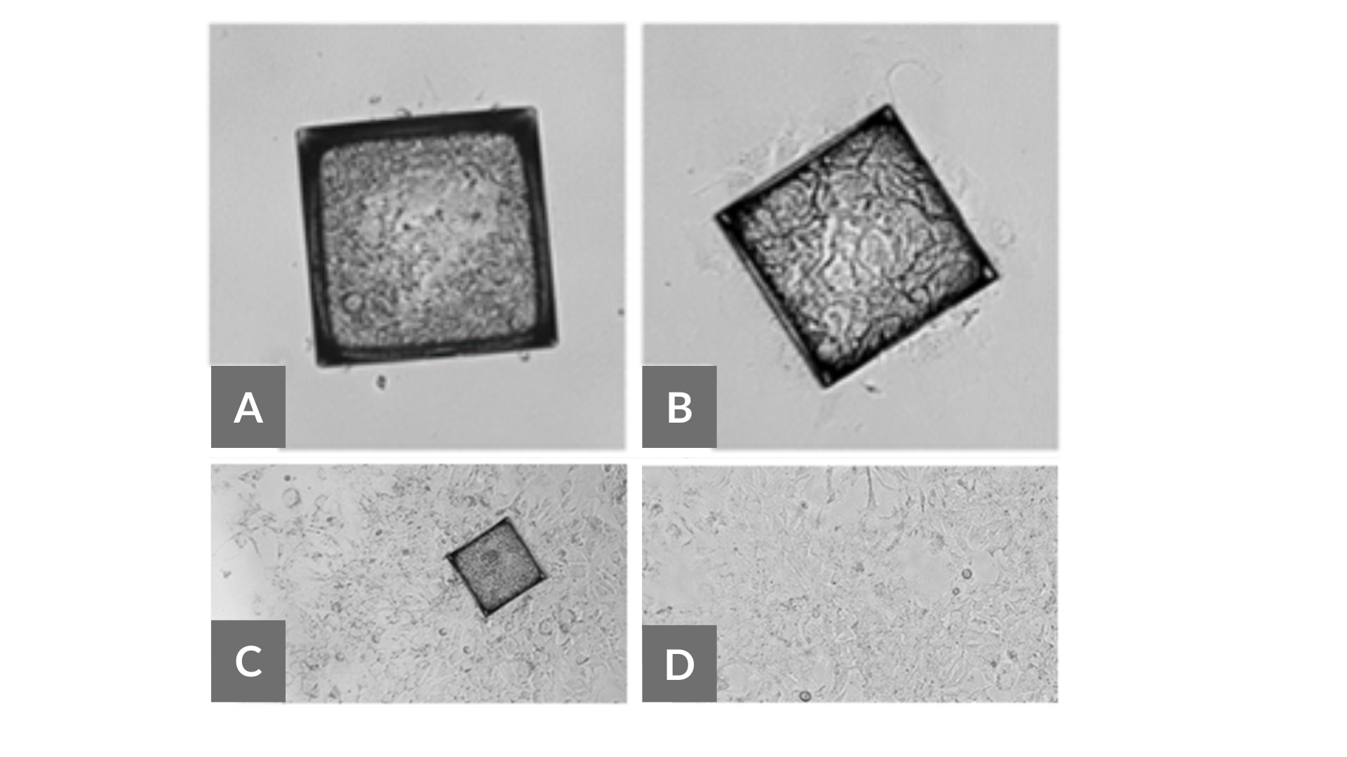
Hep3B clones post-isolation from 200S Array in conditioned or unconditioned culture media. A. Hep3B clone in unconditioned media 9 days post-isolation failed to expand off-Raft (20X magnification). B-D. Hep3B clone expanding off-Raft in conditioned media at 8 days post-isolation (20X magnification).
Epo fold-induction of hypoxic Hep3B clones (1% oxygen, 8 hours). Expected induction is 30-50 fold based on previous work with polyclonal Hep3B cells.
The Challenge
The researcher’s work routinely involves knocking in two or more unrelated fluorescent proteins into a single cell line by lentiviral methods. A significant challenge has been the isolation of strongly expressing all-positive clones for use as reporters.
The researcher desired a more facile approach to knocking in and isolating monoclonal colonies that would accelerate their work rather than having to re-engineer alternative construct designs.
A Success Story
Cells were delivered to Cell Microsystems expressing two fluorescent proteins. On a CellRaftA microscale polystyrene growth surface within an array used to spatially segregate cells/colonies while maintaining shared media access, enabling imaging over time and targeted isolation. More®Array, monoclonal colonies that were double positive for the two fluorescent markers formed and their intensity was analyzed using CellRaftA microscale polystyrene growth surface within an array used to spatially segregate cells/colonies while maintaining shared media access, enabling imaging over time and targeted isolation. More® Cytometry software. 26 monoclonal colonies were isolated and expanded before sending cells back to the researcher in less than 4 weeks.
Colonies were then verified by genomic PCR and FACS and confirmed to have the desired edit with fluorescence simplifying the researcher’s experiments and workflow.
The CellRaft ArrayConsumables containing thousands of microwells (containing CellRafts) for spatial segregation, imaging, and isolation of single cells, monoclonal colonies, or 3D cultures. More was imaged on the CellRaftA microscale polystyrene growth surface within an array used to spatially segregate cells/colonies while maintaining shared media access, enabling imaging over time and targeted isolation. More AIR® System 4 hours post-seeding to identify single cells and every subsequent 24 hours to capture clonal growth. Monoclonal colonies were identified using CellRaftA microscale polystyrene growth surface within an array used to spatially segregate cells/colonies while maintaining shared media access, enabling imaging over time and targeted isolation. More Cytometry and then isolated on day 3. Growth off-raft was imaged on a bench microscope 22 days post-isolation.
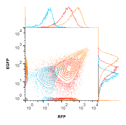
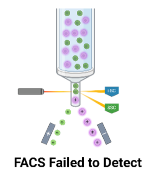
Flow cytometric analysis of THP-1 cells that have been transduced with a lentiviral construct expressing two fluorescent proteins (red) and selected (orange). Untransduced control is in blue. Contours are shown at 10% intervals with outliers as dots. All presented fluorescence has been compensated using single-color controls.
The Challenge
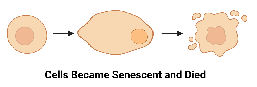
The researcher aimed to generate chemo-resistant monoclonal cancer lines to help understand intrinsic and acquired resistant mechanisms. Their standard protocol for obtaining clones that were chemo-resistant compared to their parental lines was not successful in generating clones from two cell lines of interest. Instead, cells stopped growing and went into senescence under limiting dilutionA statistical cloning approach where cells are diluted to low density to “hopefully” seed one cell per well; commonly used but can be low-efficiency for difficult-to-clone lines. More conditions.
They turned to Cell Microsystems to increase their chances of cloning those two difficult cell lines.
A Success Story
Cancer cells that were prone to senescence under stress were delivered to Cell Microsystems. Cells were seeded on a CellRaftA microscale polystyrene growth surface within an array used to spatially segregate cells/colonies while maintaining shared media access, enabling imaging over time and targeted isolation. More® Array where they shared a contiguous media volume that increased their viability. Hundreds of monoclonal colonies were screened using CellRaftA microscale polystyrene growth surface within an array used to spatially segregate cells/colonies while maintaining shared media access, enabling imaging over time and targeted isolation. More® Cytometry software which increased the likelihood of isolating cells with their desired phenotype. 43 clones in total were isolated and provided to the researcher in under 3 weeks’ time with less than one hour of hands-on time per cell line.
From the provided clones that the researcher was able to identify, 14-63% were still chemo-resistant after expansion, with 15 resistant clones for Line 1/Drug A, 5 for Line 1/Drug B, 12 for Line 2/Drug A, and 12 for Line 2/Drug B following RNA-Seq to identify chemo-resistant mechanisms.
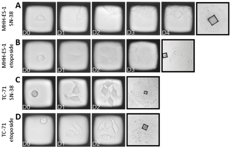
Representative time-course images of Ewing’s sarcoma cells on CellRafts (20X magnification) and outgrowth off-raft (10X magnification). CellRaftA microscale polystyrene growth surface within an array used to spatially segregate cells/colonies while maintaining shared media access, enabling imaging over time and targeted isolation. More Arrays were imaged 4 hours post-seeding to identify single cells and every subsequent 24 hours to capture clonal growth. Monoclonal colonies were identified using CellRaftA microscale polystyrene growth surface within an array used to spatially segregate cells/colonies while maintaining shared media access, enabling imaging over time and targeted isolation. More Cytometry. Growth off-raft was imaged on a bench microscope between 5-10 days post-isolation. A. MHH-ES-1 selected with SN-38, B. MHH-ES-1 selected with etoposide, C. TC-71 selected with SN-38, and D. TC-71 selected with etoposide.
The Challenge
A Success Story
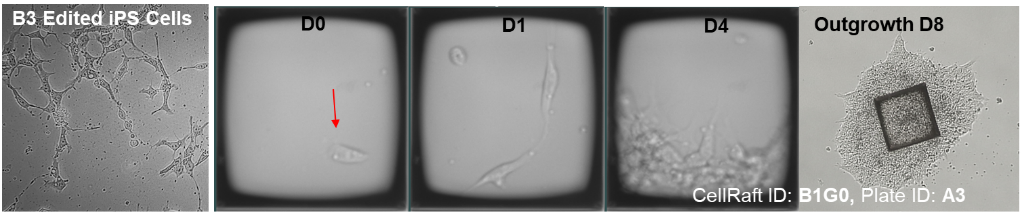
Parental iPSCs and CRISPR constructs were brought to Cell Microsystems where cells were electroporated and seeded directly on an Array. We delivered 20 polyclonal pools of edited cells to the researcher, and they confirmed 2 pools were positive. Positive cells were re-seeded on a new Array and nearly 80 clones were delivered to the researcher after 4 months of total work.
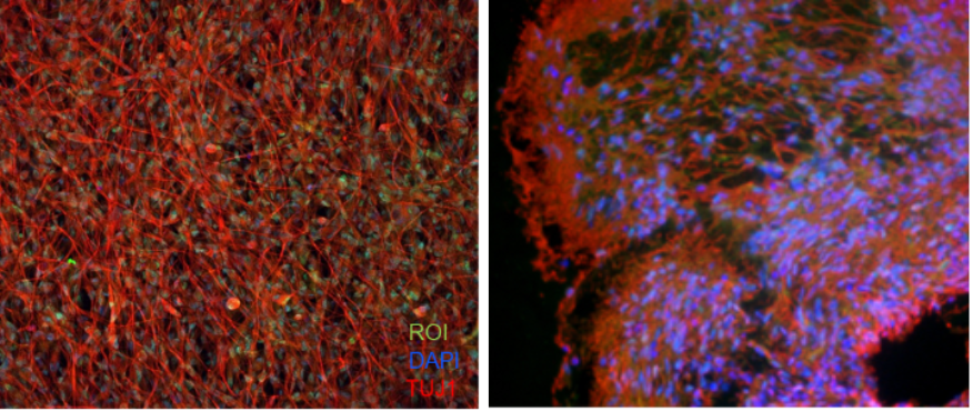
The researcher then screened 17 of the colonies and found all to be positive for their desired edit, indicating successful tagging of an elusive gene. Cells were later differentiated into 2D forebrain neurons and 3D cerebral organoids, opening new avenues for understanding its expression throughout development and providing a cell line that may be useful for high-throughput drug screening.
Attempt 1: Time course images of a representative CellRaftA microscale polystyrene growth surface within an array used to spatially segregate cells/colonies while maintaining shared media access, enabling imaging over time and targeted isolation. More containing polyclonal colonies that were isolated and screened for an expected band of interest.
Attempt 2: Positive polyclonal colonies identified in attempt 1 were expanded and seeded on a new Array, where representative CellRafts containing monoclonal colonies were isolated and outgrew in the 96-well plate.
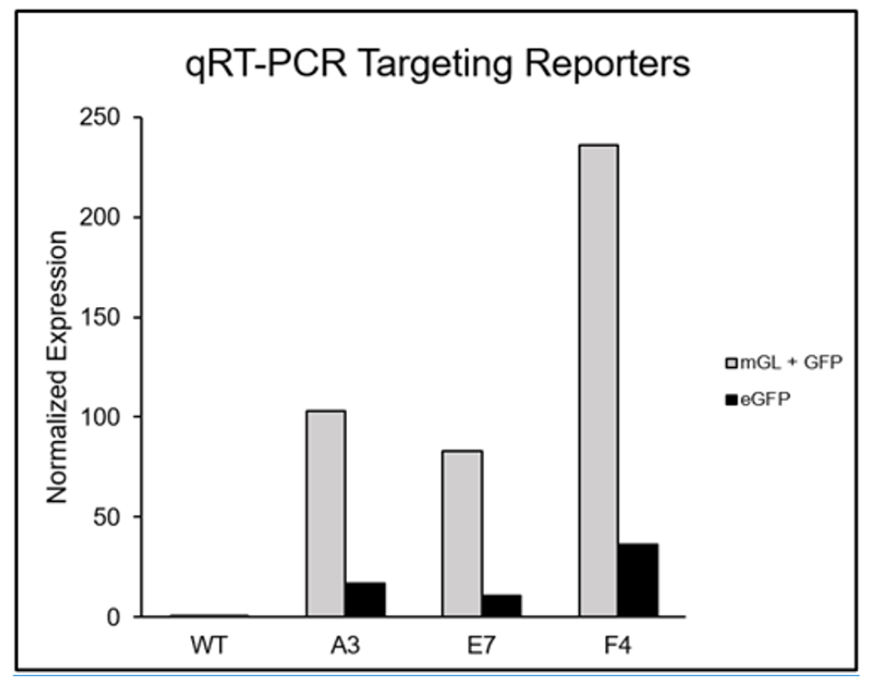
RT-qPCR of positive clones displaying GFP expression normalized to the wildtype (WT) colony.
(Left) ICC enhanced edited 11-week-old forebrain neurons for reporter of interest (ROI) (green), TUJ1 (red), DAPI (blue). Differentiation followed STEMDiff Forebrain Neuronal Kit and replating neurons for imaging followed the JOVE Protocol (Calabrese, 2019). (Right) ICC enhanced edited cerebral organoids for reporter of interest (ROI) (green), TUJ1 (red), and DAPI (blue). Differentiated using the STEMDiff Cerebral Organoid Kit.