Genomics

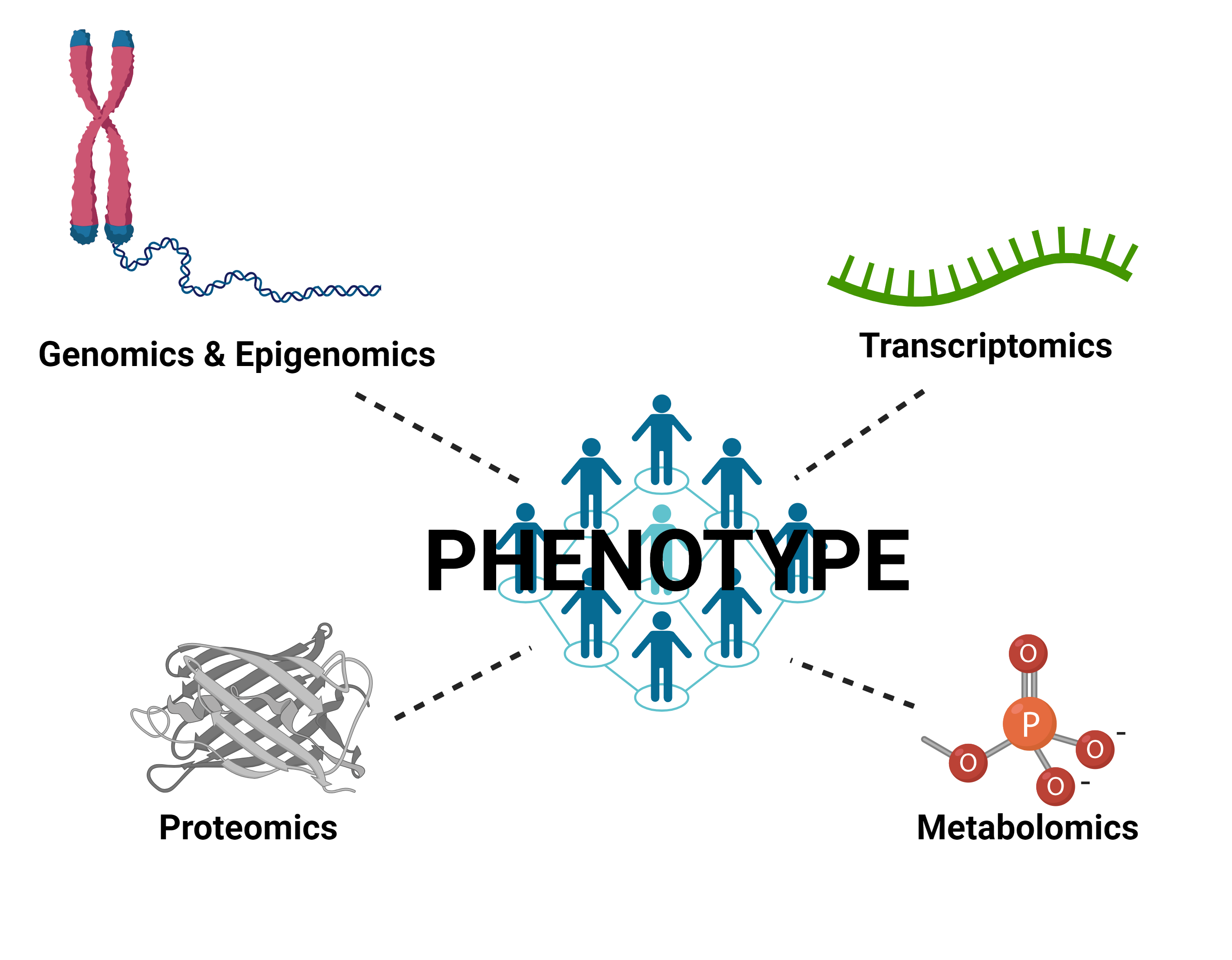
Connect Phenotype to Genotype

CellRaft AIR connecting cells to DNA editing

The CellRaft AIR System’s unique design allows users to connect downstream nucleic and protein profiles to a specific individual cell, providing greater value to each experiment.

vline gray 1

Know Exactly What Cells Are in Your Library

With typical genomic experiments, even an enriched cell population can contain undesired cells, dead cells, doublets, debris, or other artifacts. The imaging capabilities of the CellRaft AIR System provide the user with the ability to identify single cells and use software-guided tools to select desired cells based on viability or phenotypic criteria immediately after visualization.

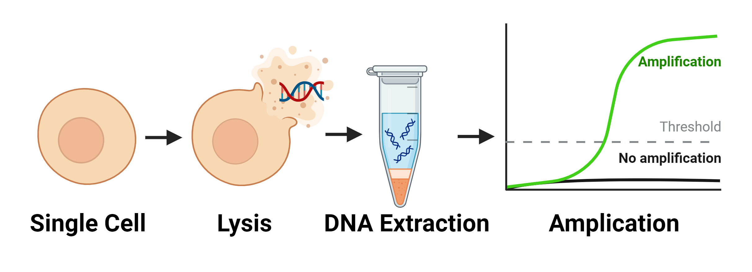
Fast, effective and gentle automated isolation helps maintain native expression profiles. Selected cells are moved into lysis buffer quickly and gently with no need for trypsin, so phenotypes remain unperturbed, and quality remains high.  No minimum required input and gentle movement allows the use of many cell types.

  • Track thousands of cells simultaneously in one consumable
  • Visualize cell stages and mutations
  • Identify biomarkers using live staining
  • Reduce the chances of doublets compared to fluorescence-activated nuclear sorting
  • Improve the quality of sensitive samples
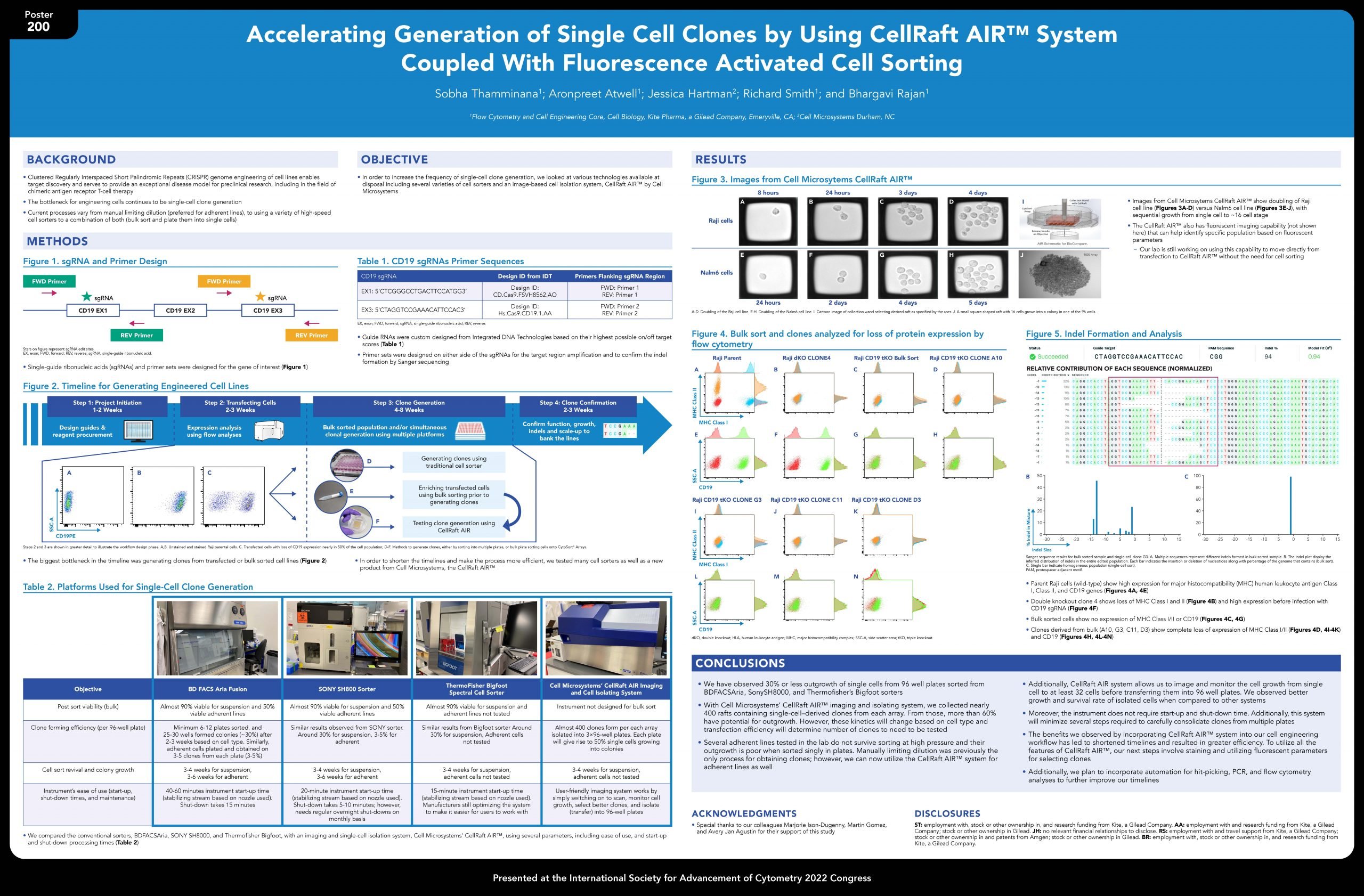
Poster Accelerating Generation of Single Cell Clones scaled

Accelerating Generation of Single Cell Clones

[glossary]A Comparison of Technologies[/glossary]
Functional Genomics

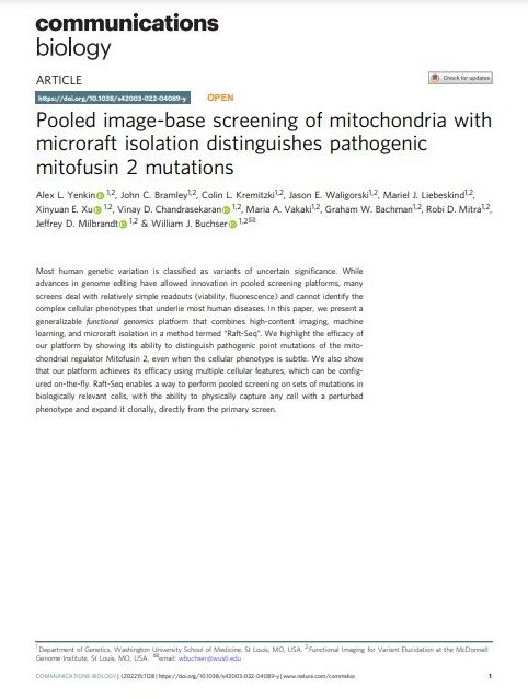
Pooled image-base screening of mitochondria with microraft isolation distinguishes pathogenic mitofusin 2 mutations

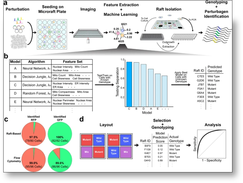
This article presents a generalizable functional genomics platform combining high-content imaging, machine learning, and CellRaft isolation termed “Raft-Seq.” This user isolates CellRafts containing cells with unique phenotypic differences allowing genotype and phenotype to be linked. In this study, Mitofusion 2 mutations were able to be distinguished efficiently.

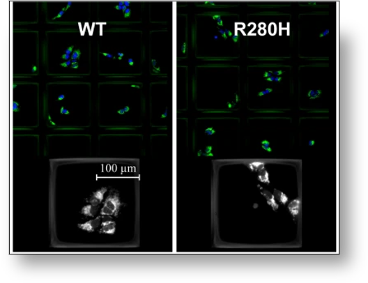
Phenotype of mutant vs. WT
CellRafts containing phenotypic differences in WT vs mutant cells
Raft-seq workflow
Raft-Seq Experimental Workflow

Article quotes about CellRaft Technology

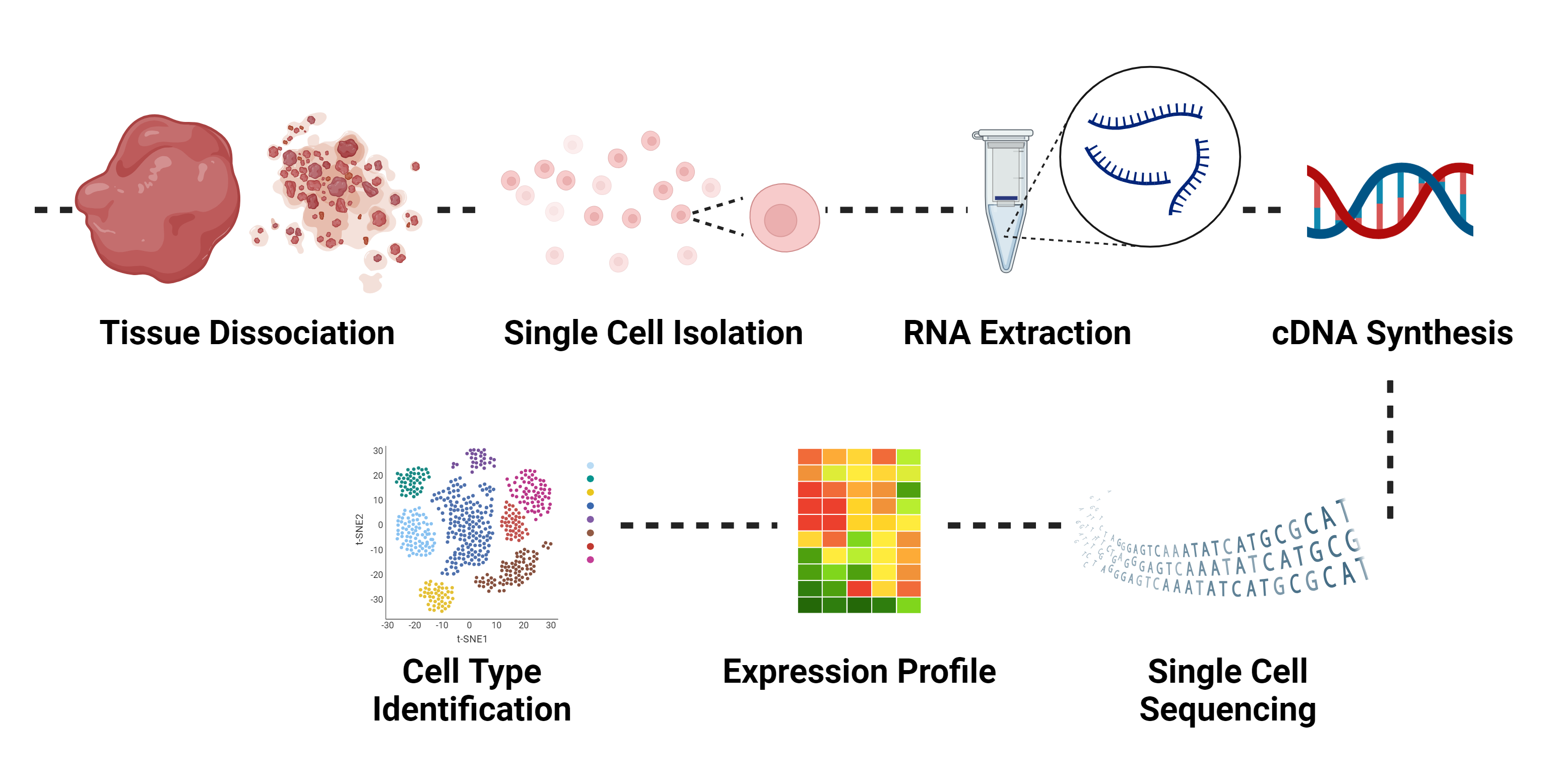
Transcriptomics and scRNA-seq

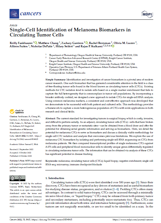
Single-Cell Identification of Melanoma Biomarkers in Circulating Tumor Cells

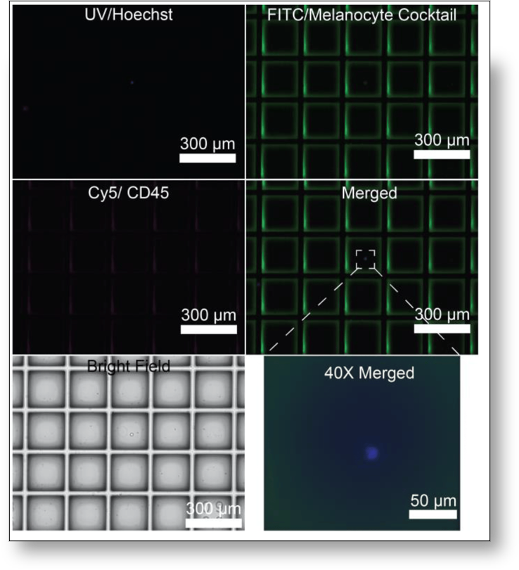
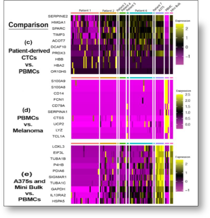
This article presents a new approach to isolate circulating tumor cells (CTCs) from a more heterogenous population using the CellRaft Array for used with both patient and cultured cells. Using a broader antibody cocktail, CTCs were first live-stained using direct immunofluorescence, seeded on an array, and then isolated for single-cell RNA analysis. Using the CellRaft Array only specific melanoma CTCs were isolated from the surrounding population demonstrating accuracy and precision of this technology.

Stained melanoma CTCs CellRaft
CellRafts containing stained melanoma CTCs for isolation
heatmaps for gene comparison CellRaft
Heatmaps of top 10 most differentially expressed gene comparisons

Article quotes about CellRaft Technology

Whole-genome Amplification

Single-cell sequencing of the small and AT-skewed genome of malaria parasites

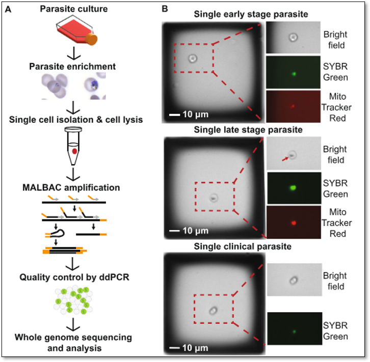
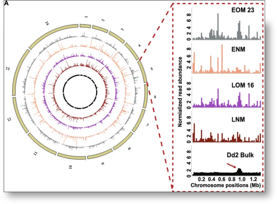
This article focused on understanding more about a life-threatening disease, malaria, which is caused from specific parasites. Using the CellRaft Array, this user was able to scan the Array on the CellRaft AIR System to identify stage specific phenotypes of single cells infected with parasites using SYBR green and Mitotracker Red staining to visualize parasite DNA and mitochondrion. This visualization allowed for the user to identify CellRafts of interest and isolate specific ones containing single infected cells for whole genome amplification later identifying SNPs and CNVs.

P falciparum infected erythrocytes CellRaft
CellRafts containing single P. falciparum-infected erythrocytes
Read abundance CellRaft
Normalized read abundance across the genome

Article quotes about CellRaft Technology

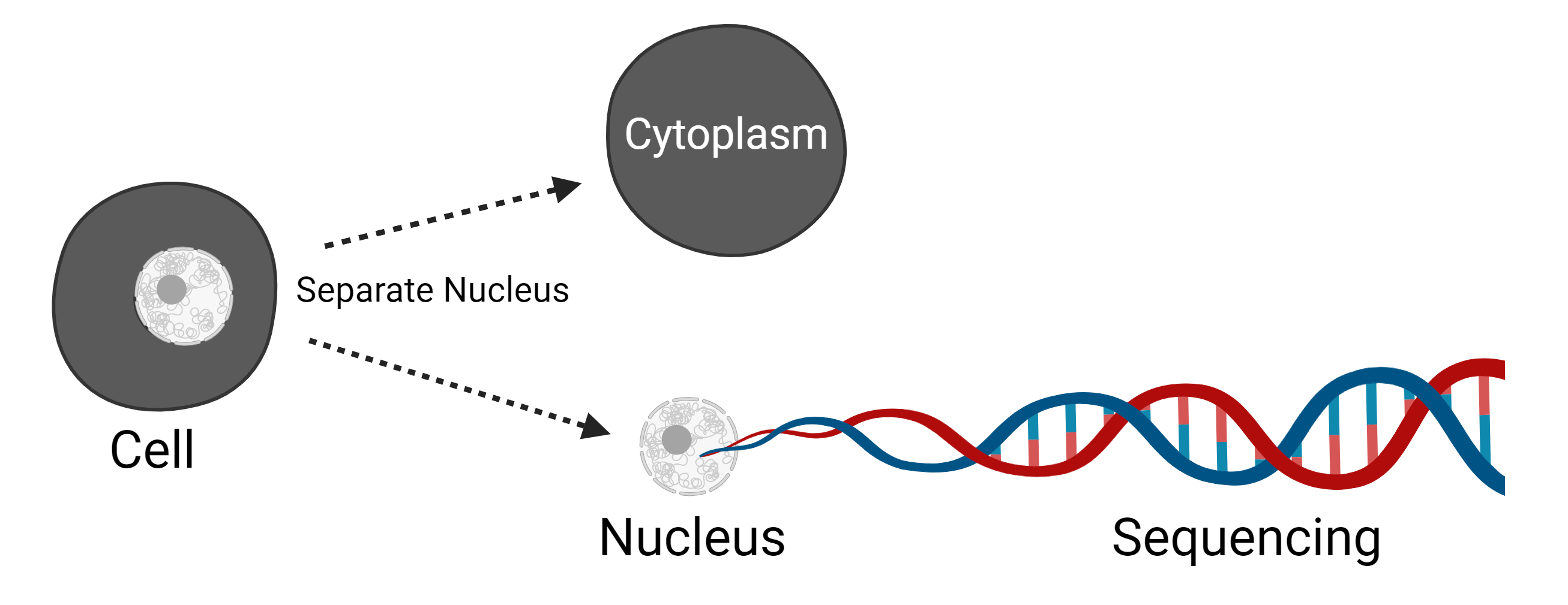
Single Nucleus Sequencing

Analyzing somatic mutations by single-cell whole-genome sequencing

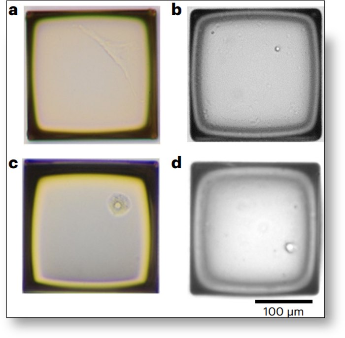
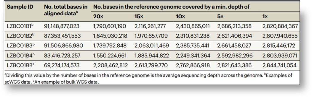
This user has expanded their single-cell isolation from originally using only adherent fibroblast cells to now other cell types including lymphocytes, hepatocytes, and nuclei for single cell isolation and sequencing. This flexibility demonstrates CellRaft Technology’s ability to be compatible with diverse cell types. Additionally, using the AIR System allowed for visualization of cells prior to sequencing while avoiding substantial damage involved in previous work with FACS.

fibroblast lymphocyte hepatocyte CellRaft
Examples of single cells or nuclei, (A) A fibroblast, (B) A lymphocyte, (C) A hepatocyte, (D) A neuronal nucleus
fractions of genome CellRaft
Fractions of single-cell genome and bulk genome covered with a sequencing depth of at least 20x

Article quotes about CellRaft Technology

Copy Number Variation

Investigation of somatic CNVs in brains of synucleinopathy cases using targeted SNCA analysis and single cell sequencing

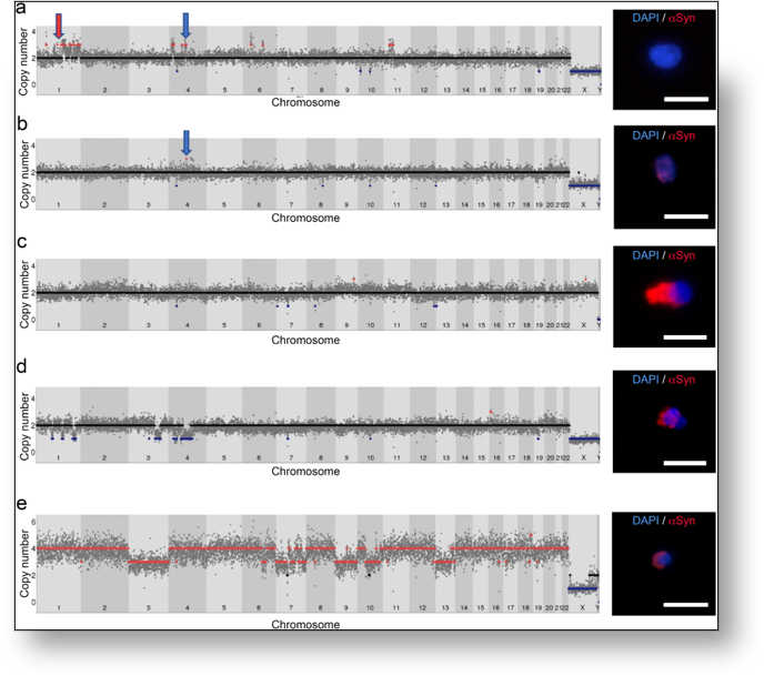
This journal focused on isolating nuclei using a CellRaft Array to evaluate somatic SNCA CNV differences for studying neurodegenerative disorders like Parkinson’s disease and multiple system atrophy. Following protocols for adhering nuclei to the Array, images were taken to obtain visual evidence of  CNVs among different nuclei after whole genome sequencing.

single nucleus enlarged nucleolus CellRaft
CellRaft containing single nucleus with enlarged nucleolus
single cell genome sequencing CellRaft
Single cell whole genome sequencing profiles showing CNVs
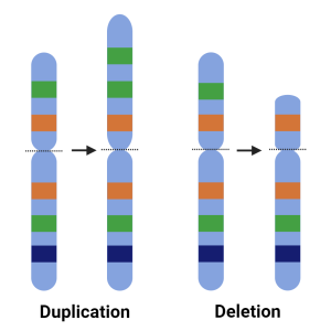
Copy number variation white background

Article quotes about CellRaft Technology

Functional Genomics

Accurate identification of single-nucleotide variants in whole-genome-amplified single cells

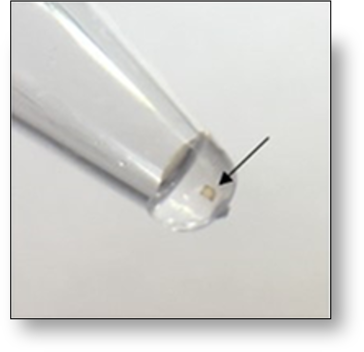
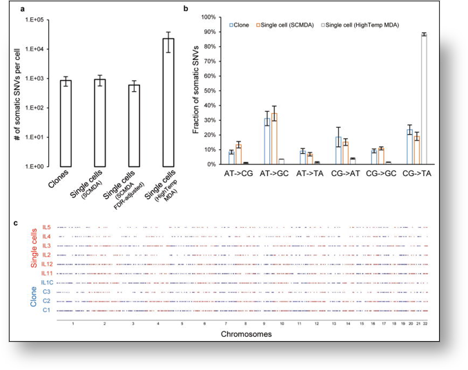
This journal highlighted the flexibility of CellRaft Technology for multiple applications. In this article, the user isolated single cells as well as monoclonal colonies using the same instrument to evaluate SNVs in both single cells and clonal colonies. With the flexibility of the AIR System, users could isolate cells in both PCR tubes and 96-well plates for their downstream assay of choice. CellRaft Technology aided this researcher in demonstrating a better solution for genome-wide DNA mutation analysis in single cells.

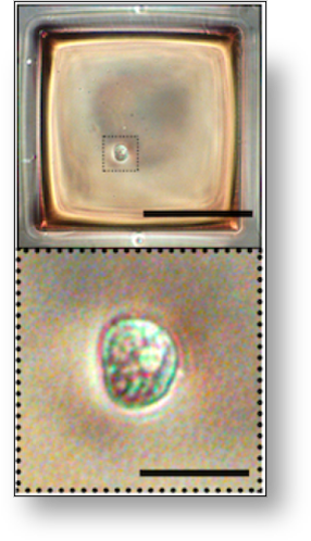
CellRaft in Pcr tube
CellRaft containing single nucleus with enlarged nucleolus
somatic SNVs CellRaft
Frequency, spectrum, and distribution of somatic SNVs
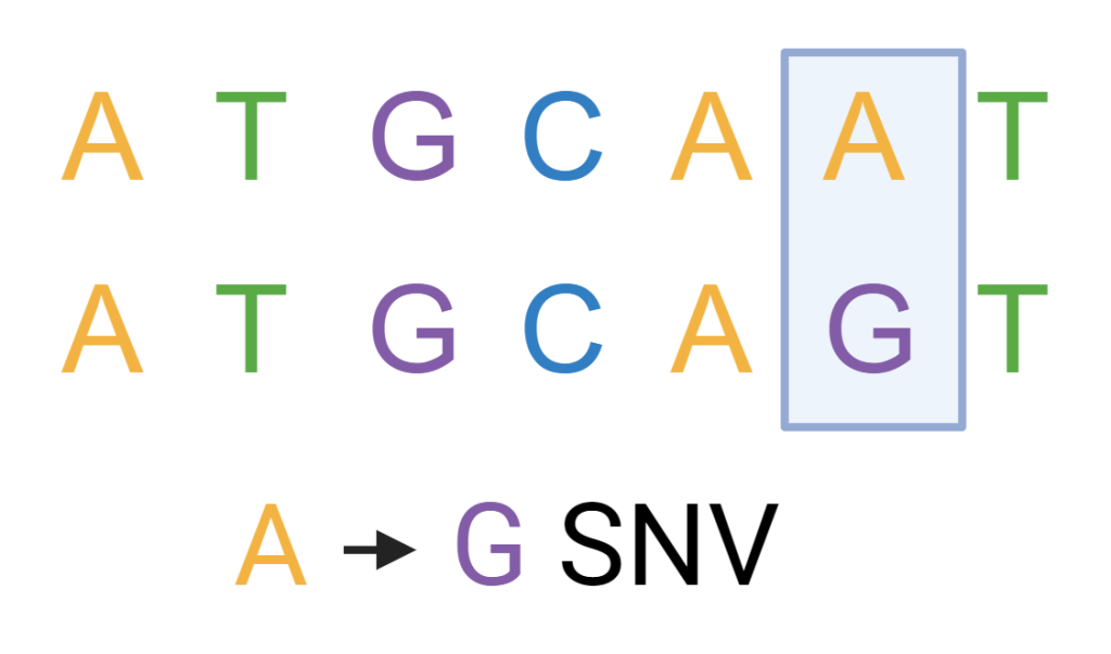
single nucleotide variation white background

Article quotes about CellRaft Technology

vline gray 1

Accelerate the Path to Discovery

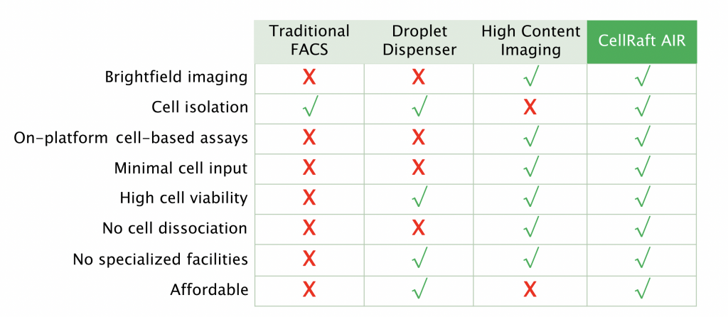
CMS versus Competitors Table
vline gray 1

How It Works

1937929574 783dc7a2c6192d3399c9cdbacb9eddeb7c1260120c5e2ba66b945d2b4bc11254 d 840?region=us

Image Cells

[glossary]Cells are plated on a CellRaft® Array using standard plating techniques, eliminating the need for trypsin, scraping, high-pressure fluidics, and limiting dilution. The array is scanned, imaged, and recorded, allowing the user to track cells as they grow over time. [/glossary]

Grow Cells

[glossary]Target cells grow unperturbed in optimized conditions in the imaging platform, increasing cell viability, and downstream read depth. The CellRaft Arrays allow different media, compound dosing, and growth conditions to be tested on a single consumablepropriety culture dish. [/glossary]

Isolate Cells

[glossary]When target cells are ready for collection, the gentle release mechanism leaves cells unperturbed, so transfer to a PCR or culture plate won’t affect the quality of the cells for downstream analysis. Cells are also moved into lysis buffer quickly, preventing the degradation of genetic material in sensitive cell lines.[/glossary]