


In a recent webinar, Dr. Jessica Hartman shared her expertise in CRISPR and gene-editing workflows, offering valuable insights into the challenges and solutions for creating reliable, edited cell lines. Here’s a look at the main takeaways and how innovative technology is changing the game for scientists in this field.
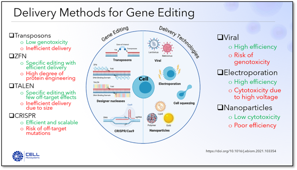
Gene editing and CRISPR workflows are often hampered by inefficiencies, high costs, and low success rates. Dr. Hartman highlighted the common difficulties scientists face in isolating single clones, the stress introduced to cells during the process, and the limited success of traditional methods like limiting dilutionA statistical cloning approach where cells are diluted to low density to “hopefully” seed one cell per well; commonly used but can be low-efficiency for difficult-to-clone lines. More. She explained that while methods like CRISPR offer precision, they can lead to issues like off-target effects or significant cellular stress.
CellRaft Technology provides a streamlined approach to improve cell viability and gene-editing efficiency. Dr. Hartman detailed how this technology allows scientists to culture and monitor cells on a single platform, reducing the need for multiple instruments and lengthy protocols. By creating a favorable, “flask-like” culture environment within the CellRaft ArrayConsumables containing thousands of microwells (containing CellRafts) for spatial segregation, imaging, and isolation of single cells, monoclonal colonies, or 3D cultures. More, researchers can maintain cell health, even for rare or challenging cell lines.
This technology offers several key advantages:
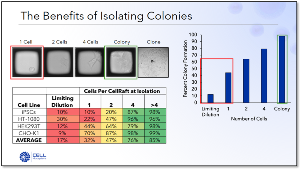
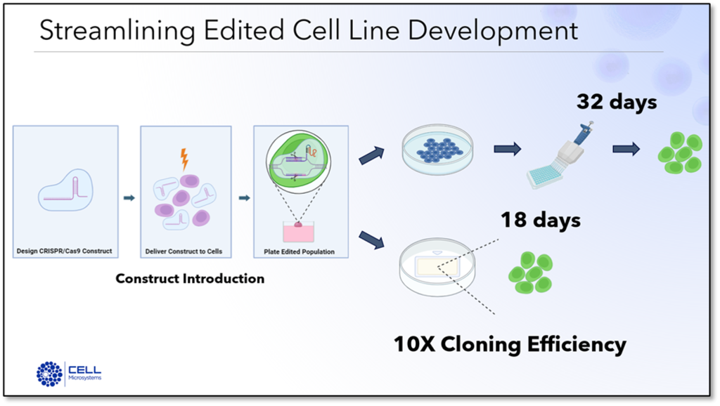
The webinar included compelling case studies demonstrating the impact of CellRaftA microscale polystyrene growth surface within an array used to spatially segregate cells/colonies while maintaining shared media access, enabling imaging over time and targeted isolation. More Technology across various applications. For example, researchers used the system to edit rare kidney podocytes, achieving clonal stability and confirmation of the desired genetic modifications within 18 days—something previously unattainable with conventional methods. Similarly, a collaboration with N.C. State yielded successful editing of iPSC lines for neurological research, even allowing differentiation into neurons and organoids without compromising cell health.
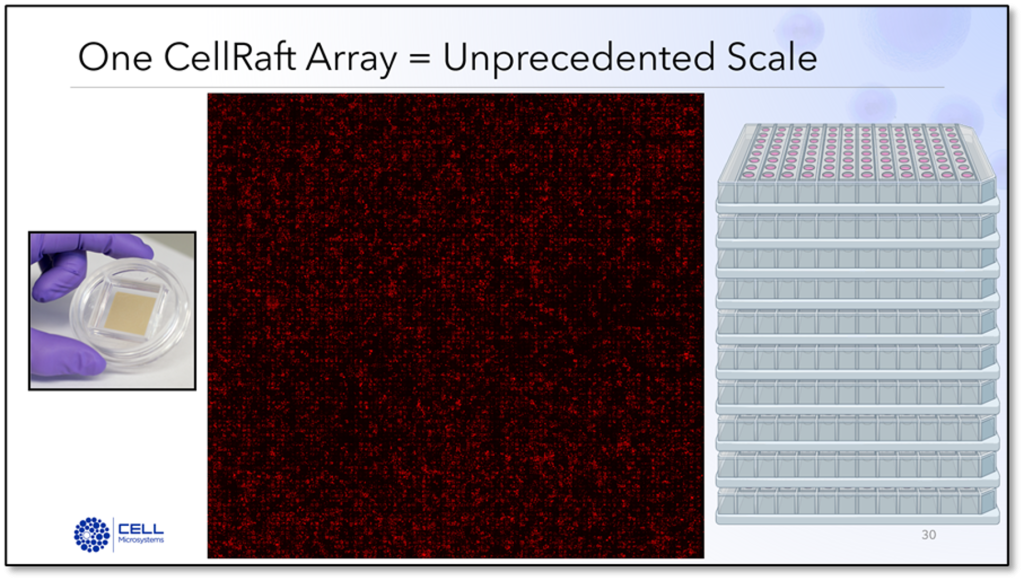
The ability of CellRaftA microscale polystyrene growth surface within an array used to spatially segregate cells/colonies while maintaining shared media access, enabling imaging over time and targeted isolation. More Technology to generate and screen thousands of clones quickly has made it a preferred choice for labs looking to conduct studies using gene edited cells. In the examples shared, researchers were able to analyze and isolate hundreds of edited cells, and easily find the edit they were looking for, reducing the need for extensive manual work.
This webinar underscored the system’s transformative potential for CRISPR research, making gene editing faster, more efficient, and ultimately more successful. For scientists tackling challenging cell types or complex editing workflows, CellRaftA microscale polystyrene growth surface within an array used to spatially segregate cells/colonies while maintaining shared media access, enabling imaging over time and targeted isolation. More Technology offers a pathway to better research outcomes and reliable, reproducible results.
If you have questions about CellRaftA microscale polystyrene growth surface within an array used to spatially segregate cells/colonies while maintaining shared media access, enabling imaging over time and targeted isolation. More Technology for CRISPR applications or are interested in our lab services, send me an email at jessica.hartman@cellmicrosystems.com.

Dr. Jessica Hartman has a B.S in Biology from the University of Virginia, a Ph.D. from Duke University in Molecular Cancer Biology and postdoctoral training in Biochemistry and Cancer Biology at Baylor College of Medicine and Duke University, respectively. She has previously served in Director-level roles, managing bioscience research and development for biotechnology companies. At Cell Microsystems, Dr. Hartman’s role is to lead the development of new and streamlined workflows using the CellRaft Technology and its associated products.


Fastest growth.
Biggest colonies.
Healthiest clones.
Prettiest morphology.
Relying on these commonly used, albeit biased, superlatives to make decisions about which cells are seemingly the “best” can ignore true biological phenotypes. As the story below highlights, the best clone may ultimately be the one that breaks the mold of these canonical criteria.
During a recent visit to a large pharma company that uses the CellRaft AIR System for cell line development, a dynamic user of the system shared a remarkable story about how the unique ability of the CellRaft AIR systemAn all-in-one platform for imaging, identifying, and isolating viable single cells, colonies, or organoids using CellRaft Arrays and software-guided selection with CellRaft Cytometry. More to support the viability of all cells, not just the strongest cells, allowed her to finally generate a long-awaited clone.
500 Clones, 0 Knockouts
In this experiment, the researcher was asked to make a CRISPR knockout of a gene in a human bladder carcinoma cell line. Using both FACS and the CellRaftA microscale polystyrene growth surface within an array used to spatially segregate cells/colonies while maintaining shared media access, enabling imaging over time and targeted isolation. More AIR in parallel, she performed five independent transductions and screened more than 500 clones without identifying a single homozygous knockout clone. As she laughingly admitted, she was picking “the best clones” that were growing robustly and assumed that the failure to generate a knockout clone was due to a technical issue such as electroporation efficiency or construct design.
Biological Breakthrough
After numerous failed attempts, she began to suspect that it could be a true biological phenotype and not a technical limitation. She noticed that many of the clones that survived on the CellRaft ArrayConsumables containing thousands of microwells (containing CellRafts) for spatial segregation, imaging, and isolation of single cells, monoclonal colonies, or 3D cultures. More were small and growing very slowly. In desperation, she isolated the slow-growing clones using the CellRaftA microscale polystyrene growth surface within an array used to spatially segregate cells/colonies while maintaining shared media access, enabling imaging over time and targeted isolation. More AIR, and remarkably, those “less beautiful” clones were her long-awaited triple homozygous knockouts. In contrast, none of the clones generated by FACS were knocked out for the gene of interest, likely because the loss of the gene conferred a growth disadvantage, and the cells could not survive after the stress of single-cell sorting.
Sharing the Knowledge
Amazingly, she was able to use this knowledge to accelerate not only her own research but also that of her colleagues at a sister site. When she learned that another group was making point mutation edits to the same gene in a different cell line, she advised them to leverage the CellRaftA microscale polystyrene growth surface within an array used to spatially segregate cells/colonies while maintaining shared media access, enabling imaging over time and targeted isolation. More Cytometry software to select CellRafts containing slow-growing, small colonies. Using this information, the group was able to generate their desired clonal cell line in ONE round of screening, not five, in only two months as compared to the year she spent uncovering the phenomenology of the gene.
From Zero to Hero
As she told me “I was a sorting devotee, but I am converted to the CellRaftA microscale polystyrene growth surface within an array used to spatially segregate cells/colonies while maintaining shared media access, enabling imaging over time and targeted isolation. More. Here, I have pretty much every well growing because we already picked the cells that expanded from the raft. With flow, we are just putting cells in a well and hoping. I’m not even turning on the cytometer anymore because there’s no point; it takes too long and isn’t going to work. The CellRaftA microscale polystyrene growth surface within an array used to spatially segregate cells/colonies while maintaining shared media access, enabling imaging over time and targeted isolation. More AIR is really improving our success rate, because we need to understand the biology, and we can use the (CellRaftA microscale polystyrene growth surface within an array used to spatially segregate cells/colonies while maintaining shared media access, enabling imaging over time and targeted isolation. More) AIR to aid in the process.”
In an increasingly fast-paced scientific landscape where “-est” is everything, this real-world experience acutely highlights the importance of allowing biology to drive discovery, not superlatives.
Click below to learn how tools like the CellRaftA microscale polystyrene growth surface within an array used to spatially segregate cells/colonies while maintaining shared media access, enabling imaging over time and targeted isolation. More AIR can make that goal easily attainable.