


Blog

Challenges and Innovations in Cell Culture: Growing the Most Difficult Cell Lines in History

Sven Bocklandt, Ph.D. Species Director and Anthony Mastracci IV, Associate Researcher from Colossal Biosciences recently joined Cell Microsystems for a webinar titled “Reviving the past, safeguarding the future: challenges and innovations in cell culture.”

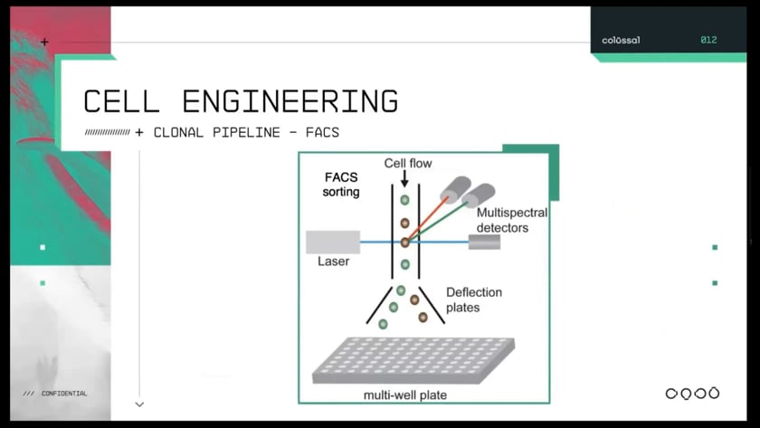
In many labs across the world, fluorescence-activated cell sorters (FACS) have remained the gold standard instrumentation for cell line development and gene editing workflows. While FACS is perhaps the most well-established automated methodology, cell sorters can present technical challenges such as:

  • The need for a dedicated highly skilled operator
  • Cross-contamination between experiments
  • Lengthy run times, which can lead to significant financial burdens and bottlenecks


In addition, for many difficult-to-culture or sensitive cell types, high-pressure fluidics-based sorting can lead to loss of cell viability, phenotypic drift, and the inability to recover rare cells.

Faced with some of these changes, Colossal Biosciences looked for an alternative technology that would augment their flow-based workflows and alleviate these pain points. Colossal discovered CellRaft Technology, a gentle and cost-effective solution for their issues.

In this webinar, Colossal scientists describe the changes to their workflow upon integrating CellRaft Technology and discuss new avenues that were not previously attainable with FACS. Take a look below as Dr. Sven Bocklandt and Anthony Mastracci IV explain how CellRaft Technology automated and instilled confidence in their workflows.

1882480091 a62e95588fba2cadc265eba7aae2a3aeec0b8e43c2546176fb0b5fe56de000c7 d 840?region=us

How does CellRaft Technology isolate cells more gently than FAC sorting?

FACS

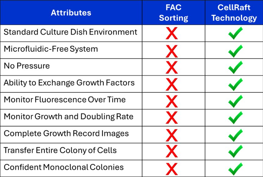
To isolate cells to make monoclonal cell lines, the standard in the field really is to use FACS or fluorescence activated cell sorting. That puts several stresses on these cells. They sit in a sorting buffer sometimes for a long time, they're exposed to high pressure during the sorting, and then importantly they end up alone in a well without the ability to secrete and share growth factors with other cells.

CellRaft AIR

We are using the CellRaft AIR, from Cell Microsystems to isolate cells in a way that is more gentle than FAC sorting. We seed cells onto a culture plate that is made up of thousands of microscopic rafts where they share the media and thus they can keep exchanging growth factors as if they are in a standard cell culture dish. We were concerned about cells transferring between wells, but we’ve been able to confirm the cells maintain monoclonality.

logo crisper black
Anthony Mastracci IV
Associate Researcher
1882536707 9e9b4cdb98ffa121e0a0aef16e7eb0fa1a9b324577b9ee27b367bdf5733c7073 d 840?region=us

How did CellRaft Technology automate Colossal’s manual workflow?

FACS

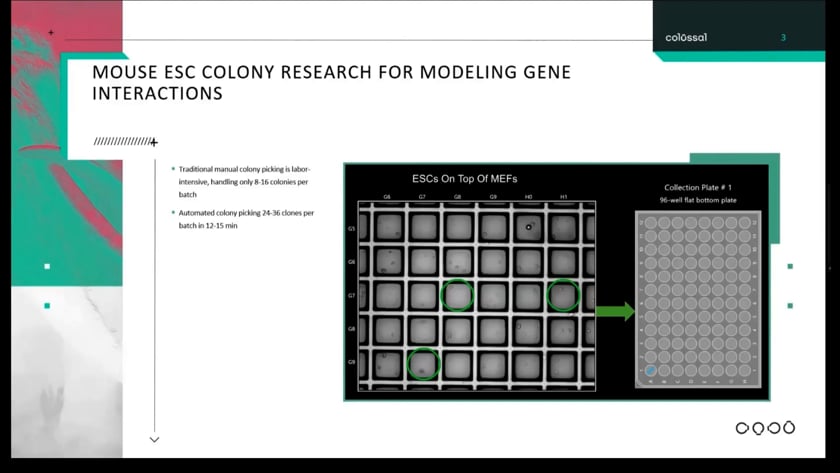
Cloning mouse embryonic stem cells is a tedious process of spending hours hunched over a stereoscope, meticulously using a P20 pipette to manually pick clones from a 6-well flask or larger. It is quite labor-intensive and places a significate strain on our scientists, consequently diverting their attention from higher-level tasks.

CellRaft AIR

Upon implementing the CellRaft, we immediately noticed its ability to automatically pick clones of interest. This, in turn, freed up our scientists to focus on other lab tasks. Now the ability to observe individual ESLs just four hours after seeding expands our editing workflows and instils greater confidence in our research. This enables us to explore new avenues that were previously inaccessible.

logo crisper black
Anthony Mastracci IV
Associate Researcher
1882561128 d0f9e218e117e9debe1f7290b125faba84df38d57ee08ba5d9b17b694ab62570 d 840?region=us

How does CellRaft Technology ensure monoclonality?

FACS

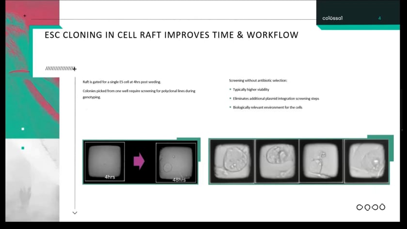
Our approach involved seeding ESLs at low density on a six-well plate, followed by waiting seven to nine days before examining under the stereoscope. This method occasionally resulted in the formation of polyclonal lines when two ESLs either landed in close proximity to one another or failed to fully dissociate appearing as a single colony days later. Consequently, screening for polyclonal during genotyping became necessary.

CellRaft AIR

Now using CellRaft Technology, we have the ability to visualize single ES cells just four hours post-seeding possessing greater confidence in the monoclonal nature of colonies being generated. And as matter of fact, since implementing the CellRaft, we have yet to encounter a polyclonal ES colony.

logo crisper black
Anthony Mastracci IV
Associate Researcher
1883381254 bbf29503e89d622691b9edd22f93e346c24ef1f723d0c6a1fcaa79d4ec5d1a89 d 840?region=us

How does CellRaft Technology aid in gene editing workflows?

CellRaft AIR

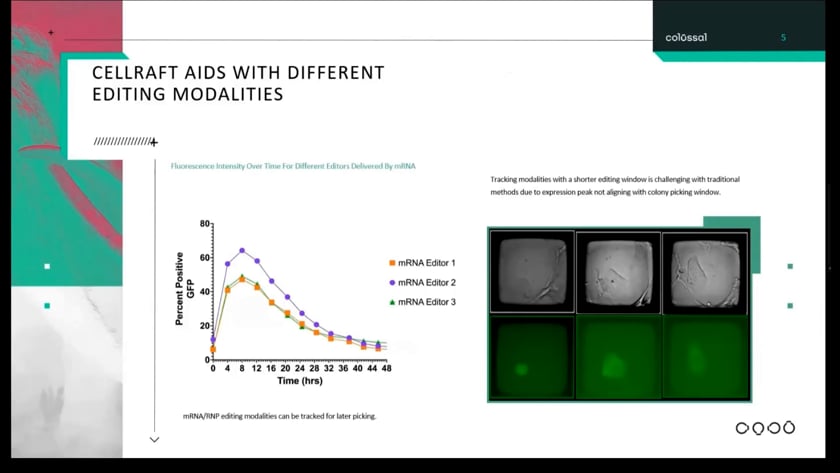
mRNA has a short editing window peaking at around eight hours and tapering off over the course of 48 hours visualizing early ESL transfections. We can see the tapering off of the fluorescence begining to fade resulting in low SNR. This now demonstrates to us that we can start using fluorescence for positive selection for mouse ESLs.

logo crisper black
Anthony Mastracci IV
Associate Researcher
Colossal Blog

Upon implementing CellRaft Technology, Colossal automated their clone picking, noticed cells were more viable within their natural environment, and could visualize earlier within the short editing window. They were able to avoid excessive costly screening, having confirmed monoclonal colonies in their hands. These advantages demonstrate the flexibility, ease of use, and confidence Colossal has in the CellRaft Technology. Colossal now has the ability to accelerate their research freeing up scientists from constantly sorting, re-analyzing, or additionally screening their cells.

To see more information on the technology that helped Colossal accelerate their workflows and enable their scientists to focus their attention back to higher-level tasks, visit our website and read about CellRaft Technology.

Lexi Land B.S. 300x300 1
Lexi Land, B.S.
Research Associate II | lland@cellmicrosystems.com

Lexi Land contributes to developing scientific workflows that are compatible with the CellRaft® AIR System. Her focus at Cell Microsystems involves stem cell research, 3D organoid work, as well as adherent and suspension cell culture. Lexi received a Bachelor of Science in Biological Sciences from North Carolina State University with a focus in molecular, cellular, and developmental biology.

Related Posts

Unlocking the Secrets of Antibiotic Resistance: How Shear Flow Systems Are Revolutionizing the Fight Against Superbugs

In the ongoing battle against antibiotic-resistant bacteria, or “superbugs,” scientists are constantly seeking innovative tools to understand the mechanisms of resistance and develop effective treatments. One such groundbreaking technology is shear flow systems, which have emerged as game-changers in microbiological research. By simulating the dynamic conditions of bacteria’s natural environments, such as the human body or food processing, shear flow systems provide unique insights into bacterial behavior and resistance mechanisms that traditional static methods simply cannot achieve. In this blog post, we’ll explore the benefits of using shear flow systems to combat antibiotic resistance and develop next-generation therapies.

Why Shear Flow Systems Are Essential for Studying Antibiotic Resistance

Antibiotic resistance is a complex phenomenon influenced by a variety of factors, including genetic mutations, horizontal gene transfer, and the formation of biofilms. Traditional laboratory methods often fail to capture the dynamic nature of these processes, leading to incomplete or misleading results. Shear flow systems address this limitation by providing a more realistic environment for bacterial growth and interaction. Here’s how:

1. Mimicking Real-World Conditions

In the human body, bacteria are rarely in a static state. They are constantly exposed to fluid flow, such as blood circulation or urine flow, which influences their behavior and resistance mechanisms. Shear flow systems can replicate these conditions, allowing researchers to study how bacteria respond to antibiotics under realistic physiological conditions. This is critical for understanding how resistance develops and persists in vivo. This benefit was recently highlighted in a webinar presented by Dr. Katharina Richter, a microbiology researcher from the University of Adelaide in Australia. Using a high-throughput BioFlux Shear Flow System, Dr. Richter and her team were able to test 3 different methods of superbug treatments under physiological conditions.

2. Studying Biofilm Formation and Resistance

Biofilms—structured communities of bacteria encased in a protective matrix—are a major contributor to antibiotic resistance. Biofilms are notoriously difficult to treat because they shield bacteria from antibiotics and the immune system. Shear flow systems enable researchers to study biofilm formation in real time, observing how bacteria adhere to surfaces, form microcolonies, and develop resistance under flow conditions. For example, using a BioFlux, Dr. Richter was able to leverage high-resolution imaging to obtain unprecedented insight into the biofilm killing and prevention efficacy of a copper and diethyldithiocarbamate (DDC) combination nanoparticles¹. The group is currently investigating the use of a Cu-DDC infused gel that can be injected into wounds that are at high risk for chronic infection, such as hernia.

…
The Evolution of Polymicrobial Assessment: From Culture to Automation

The study of biofilm communities presents unique challenges in experimental design and analysis. While mixed bacterial populations offer fascinating insights into microbial interactions and community dynamics, accurately characterizing and investigating these complex systems has previously required sophisticated and tedious technical approaches. Here, we will discuss advances in technology that have helped to simplify and streamline polymicrobial investigations.

Expanding Research Assays

Throughout the last decade, a trend for growing and assaying biofilms under shear flow has gained traction among microbiologists. The rationale for investigating biofilms under flow is multi-dimensional. A recent Cell Microsystems survey asked why microbiologists are using shear flow found that the top responses were:   

  1. Improved physiological relevance
  2. Enhanced biofilm attachment and detachment
  3. Better nutrient and gas exchange

Of the scientists surveyed, 40% indicated that they use a BioFlux Shear Flow System. This system provides unique advantages over other methods to induce shear stress, such as slide-based parallel plate flow chambers and benchtop rockers.

BioFlux uses microfluidic channels embedded onto the bottom of standard-sized 6, 24, or 48-well plates to create isolated and independent flow chambers where user-defined wall shear stress is initiated and maintained by digitally controlled air pressure. This not only simplifies the setup and experimental processes but also ensures that experiments are contained and do not contaminate the shear flow system.

Although shear flow has been thoroughly demonstrated to improve the biological relevance of biofilm investigations1, an often overlooked aspect of biofilm physiology is the significant role that multicellular aggregates play in biofilm formation2. When investigating Pseudomonas aeruginosa, it has been demonstrated that multicellular aggregates lead to faster biofilm formation and enhanced resistance to antimicrobials and immune responses3. In addition, multispecies microbial aggregates have been shown to dominate single cells in human saliva4. Together, these data show that aggregates are important areas of investigation because they are more likely to represent physiological biofilm formation and growth.

Traditional Culture-Based Methods [elementor-element k="b4c7e08eabafaf9b48ea36c77ccfdf87"…
Advances in Biofilm Research: How the Experts are Overcoming Obstacles
Recently, some members of the Cell Microsystems team traveled to the United Kingdom, to speak with researchers at some of…