Historically, cell line development has predominantly relied on a limited number of cell lines such as CHO-K1, HEK293T, and HeLa. However, as biotechnology continues to advance, there is a growing demand for cell line development platforms that can support a broad range of cell types.
To demonstrate the power and versatility of CellRaftA microscale polystyrene growth surface within an array used to spatially segregate cells/colonies while maintaining shared media access, enabling imaging over time and targeted isolation. More Technology to enable clonal cell growth, our team set an ambitious goal to test and characterize 100 unique cell lines on the CellRaft ArrayConsumables containing thousands of microwells (containing CellRafts) for spatial segregation, imaging, and isolation of single cells, monoclonal colonies, or 3D cultures. More. We successfully accomplished this milestone when we added the 100th cell line in December 2022.
During this endeavor, our team gained valuable insights into the specific requirements of each individual cell line. Here are four key points we learned along the way:
Some cell lines are very difficult to clone using traditional cloning methods that place a single cell alone in a well, creating a lack of important growth factors and mitogens from neighboring cells. The CellRaft ArrayConsumables containing thousands of microwells (containing CellRafts) for spatial segregation, imaging, and isolation of single cells, monoclonal colonies, or 3D cultures. More provides a unique cloning environment where single cells can divide in a flask-like bulk culture environment and share contiguous media while maintaining spatial segregation for monoclonalityEvidence that a colony/line originated from one cell; often supported by time-course imaging and traceability. More. Single cell cloning efficiency was greater than 70% for most of the cell lines that we tested, compared to the typical 0-30% cloning efficiency of a traditional limiting dilutionA statistical cloning approach where cells are diluted to low density to “hopefully” seed one cell per well; commonly used but can be low-efficiency for difficult-to-clone lines. More workflow (Figure 1). When tested against limiting dilutionA statistical cloning approach where cells are diluted to low density to “hopefully” seed one cell per well; commonly used but can be low-efficiency for difficult-to-clone lines. More, the CellRaft ArrayConsumables containing thousands of microwells (containing CellRafts) for spatial segregation, imaging, and isolation of single cells, monoclonal colonies, or 3D cultures. More yielded hundreds more confirmed clones (Figure 2).
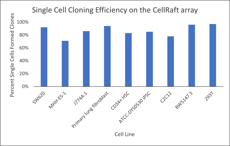 Figure 1: Single cell cloning efficiency of various cell types tested on the CellRaft ArrayConsumables containing thousands of microwells (containing CellRafts) for spatial segregation, imaging, and isolation of single cells, monoclonal colonies, or 3D cultures. More. Defined as the percentage of single cells that formed clones, single cell cloning efficiency was greater than 70% for most cells tested.
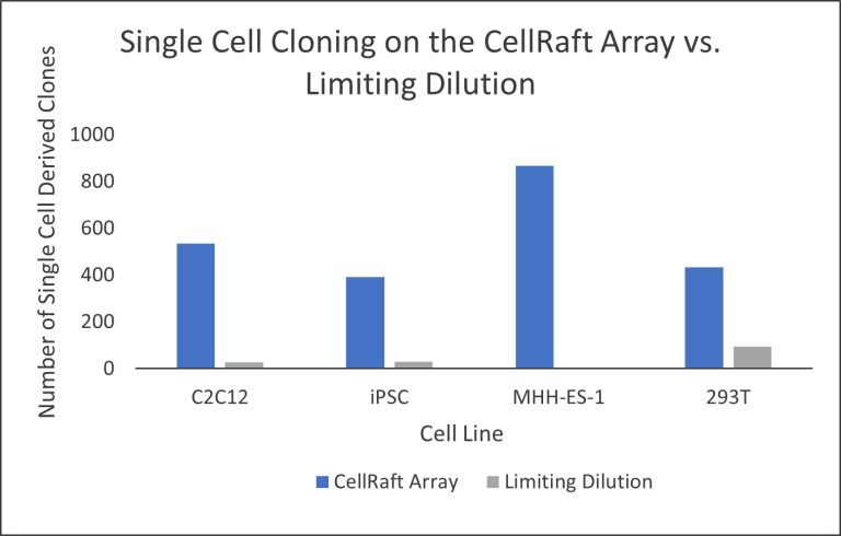
Figure 2: Total number of single cell derived clones obtained from various cell types on one CellRaft ArrayConsumables containing thousands of microwells (containing CellRafts) for spatial segregation, imaging, and isolation of single cells, monoclonal colonies, or 3D cultures. More or three limiting dilutionA statistical cloning approach where cells are diluted to low density to “hopefully” seed one cell per well; commonly used but can be low-efficiency for difficult-to-clone lines. More plates per cell line. The CellRaft ArrayConsumables containing thousands of microwells (containing CellRafts) for spatial segregation, imaging, and isolation of single cells, monoclonal colonies, or 3D cultures. More outperformed limiting dilutionA statistical cloning approach where cells are diluted to low density to “hopefully” seed one cell per well; commonly used but can be low-efficiency for difficult-to-clone lines. More by generating hundreds more single cell derived clones. MHH-ES-1 cells failed to proliferate in limiting dilutionA statistical cloning approach where cells are diluted to low density to “hopefully” seed one cell per well; commonly used but can be low-efficiency for difficult-to-clone lines. More plates but yielded 867 single cell derived clones on just one CellRaft ArrayConsumables containing thousands of microwells (containing CellRafts) for spatial segregation, imaging, and isolation of single cells, monoclonal colonies, or 3D cultures. More.
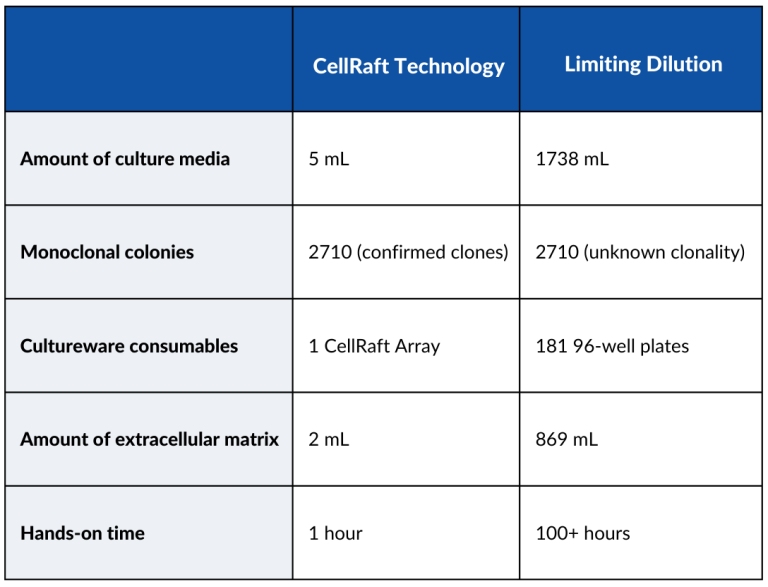
One CellRaft ArrayConsumables containing thousands of microwells (containing CellRafts) for spatial segregation, imaging, and isolation of single cells, monoclonal colonies, or 3D cultures. More can yield thousands of clones in a workflow that, for most cells, requires less than three weeks from seeding cells on the CellRaft ArrayConsumables containing thousands of microwells (containing CellRafts) for spatial segregation, imaging, and isolation of single cells, monoclonal colonies, or 3D cultures. More to a full 96-well plate of expanding clones. Renca cells seeded on a 100 μm Quad-Reservoir CellRaft ArrayConsumables containing thousands of microwells (containing CellRafts) for spatial segregation, imaging, and isolation of single cells, monoclonal colonies, or 3D cultures. More yielded 2,710 single cell derived clones after only three days of incubation.
Following the isolation of CellRafts containing monoclonal colonies to a 96-well plate, cells began to expand off-raft after only three days.
Assuming a generous limiting dilutionA statistical cloning approach where cells are diluted to low density to “hopefully” seed one cell per well; commonly used but can be low-efficiency for difficult-to-clone lines. More efficiency of 15 clones per 96-well plate, a limiting dilutionA statistical cloning approach where cells are diluted to low density to “hopefully” seed one cell per well; commonly used but can be low-efficiency for difficult-to-clone lines. More workflow to obtain a similar number of clones would require 181 96-well plates, over 1700 mL of media, and countless hours of hands-on time. If Renca cells required an extracellular matrix-like Matrigel, only 2 mL would be required to coat the CellRaft ArrayConsumables containing thousands of microwells (containing CellRafts) for spatial segregation, imaging, and isolation of single cells, monoclonal colonies, or 3D cultures. More compared to 869 mL required to coat 181 96-well plates.
Cells vary widely in morphology, preferred seeding density, and extracellular matrix requirements. While some cells like iPSCs prefer the extra growth area of the 200 μm CellRafts, other cells prefer the higher seeding density that the 100 μm CellRafts offer. Cells also range in doubling time, from less than 10 hours for BHK-21 cells to more than 40 hours for PC-12 adrenal cells, requiring modulation of imaging frequency and time on the array until isolation into a collection plate (Figure 3).
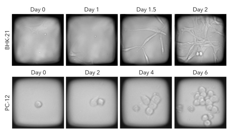
Figure 3: BHK-21 cells proliferating on a 200 μm CellRaftA microscale polystyrene growth surface within an array used to spatially segregate cells/colonies while maintaining shared media access, enabling imaging over time and targeted isolation. More and PC-12 cells proliferating on a 100 μm CellRaftA microscale polystyrene growth surface within an array used to spatially segregate cells/colonies while maintaining shared media access, enabling imaging over time and targeted isolation. More. BHK-21 cells prefer the larger 200 μm CellRaftA microscale polystyrene growth surface within an array used to spatially segregate cells/colonies while maintaining shared media access, enabling imaging over time and targeted isolation. More and require only two days on the CellRaft ArrayConsumables containing thousands of microwells (containing CellRafts) for spatial segregation, imaging, and isolation of single cells, monoclonal colonies, or 3D cultures. More before isolation to a 96-well collection plate. Slow-growing PC-12 cells prefer the smaller 100 μm CellRaftA microscale polystyrene growth surface within an array used to spatially segregate cells/colonies while maintaining shared media access, enabling imaging over time and targeted isolation. More and are allowed to proliferate for at least six days before isolation.
When initiating the culture of any cell type on an extracellular matrix, various matrix concentrations should be tested to determine an optimal concentration. Various concentrations of Matrigel, a common extracellular matrix used to coat tissue culture surfaces, were tested on the CellRaft ArrayConsumables containing thousands of microwells (containing CellRafts) for spatial segregation, imaging, and isolation of single cells, monoclonal colonies, or 3D cultures. More with a stable iPSC line. Single cell cloning efficiency increased from 49% to 74% with increasing concentrations of Matrigel (Figure 4). While cell lines typically prefer a higher extracellular matrix concentration when propagated clonally on the CellRaft ArrayConsumables containing thousands of microwells (containing CellRafts) for spatial segregation, imaging, and isolation of single cells, monoclonal colonies, or 3D cultures. More, only 2 mL of dilute matrix is required for coating while at least 14 mL of matrix is required to coat three limiting dilutionA statistical cloning approach where cells are diluted to low density to “hopefully” seed one cell per well; commonly used but can be low-efficiency for difficult-to-clone lines. More plates.
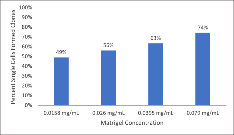 Figure 4: Single cell cloning efficiency of iPSCs on the CellRaft ArrayConsumables containing thousands of microwells (containing CellRafts) for spatial segregation, imaging, and isolation of single cells, monoclonal colonies, or 3D cultures. More with increasing concentrations of Growth Factor Reduced Matrigel (Corning).
In our endeavor to test 100 different cell lines using CellRaftA microscale polystyrene growth surface within an array used to spatially segregate cells/colonies while maintaining shared media access, enabling imaging over time and targeted isolation. More technology, we discovered that the success of each cell type relies on meticulously characterized conditions. Variations between cells influence the optimal CellRaft ArrayConsumables containing thousands of microwells (containing CellRafts) for spatial segregation, imaging, and isolation of single cells, monoclonal colonies, or 3D cultures. More size, seeding density, and extracellular matrix required to get the most out of cloning cells. One prevailing theme that emerged from our testing of 100 cell lines was that cells are much easier to clone when they can share media with neighboring cells, and happy cells make happy scientists.

Anna works on the Product Applications team where she develops novel single-cell workflows that are compatible with the CellRaft AIR systemAn all-in-one platform for imaging, identifying, and isolating viable single cells, colonies, or organoids using CellRaft Arrays and software-guided selection with CellRaft Cytometry. More. Before joining Cell Microsystems, she worked as an undergraduate research assistant in a microbiology laboratory and as an intern on a quality control team at Lonza Biologics. She holds a Bachelor of Science in Microbiology and Biochemistry from the University of Maine.