Blog

Transforming Research Challenges, Stanford Success Story

As my mother, a seasoned scientist and lab veteran who has trained countless students and post-docs, likes to say, “They don’t call it RE-search for nothing.  It’s because we RE-do experiments over and over again until it works.”  So, it should come as no surprise that she cautioned me to think long and hard about becoming a career researcher.

In the folly of my youth, I didn’t listen, and as mothers tend to be, it turns out she was completely right.  Day after day, scientists show up to the lab striving to achieve the impossible.  Despite long hours, grueling experiments, and weeks or months of failure, we persevere, hoping for that elusive breakthrough that can impact human health.  Yet sometimes, despite our best efforts, flawless experimental designs, and meticulous lab work, our tools fall short, leaving us to RE-peat our labors.

It is our keen understanding of this frustration that drove us to launch our “Clone Challenge”.  Our aim was simple: provide struggling scientists access to novel technology that could solve the bottleneck inhibiting their progress.  Our first clone challenge winner came to us from Stanford University. We were excited by the complexity of the problem and the opportunity to help.

In their application, the Stanford team explained that they were using a human glioblastoma cell line (the most malignant adult brain cancer type), which must be cultured in suspension as spheroids to prevent differentiation. However, as is often the case, the cell line is not homogenous, and approximately 90% of the population has one type of genetic amplification, while 10% of the population has a different genetic amplification of the same gene.  For months, they had been attempting to derive clonal populations of these two genotypes without success, as these cells do not grow well as single cells. Moreover, the screening necessary to confirm the genotypes was labor-intensive, expensive, and not easily scalable.

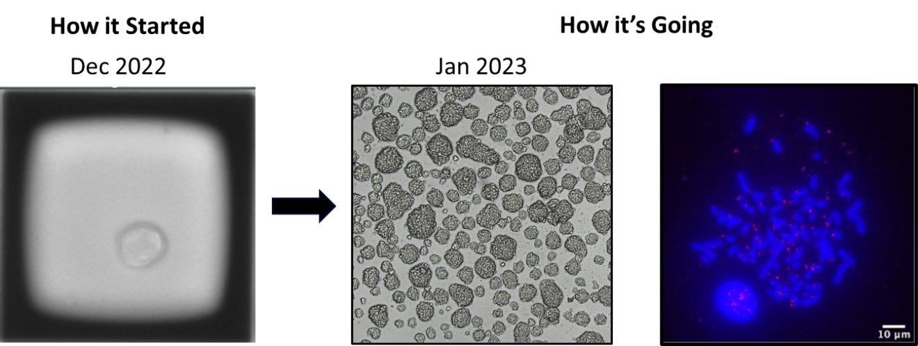
The team at Stanford provided us with their cell line, and we used CellRaft Technology to derive monoclonal cell lines from the bulk parental line. Although the initial promise of the clone challenge was five monoclonal cell lines, due to the low frequency (1 in 10) of one of the genotypes, we realized we needed to screen as many as possible.  Over the course of about six weeks, we generated clonal cell lines that maintained their ability to grow as spheroids (see below).  Upon returning the clones to Stanford, they validated that 26 of the lines were correct, and they were able to obtain isogenic matched pairs with both genotypes after confirmation with their orthogonal methods. 

As a scientist who has failed in the lab all too often, the response from the team at Stanford was profoundly gratifying:

“Thank you so much for your help earlier in helping us to get monoclonally expanded cells. We are all very happy with the outcome as it has been quite a challenging cell system for monoclonal expansion. The clones are a great addition to the lab’s expanding cell line models.”

The moral of the story?  Sometimes, you just need a little help. This is why I show up every day as a scientist at Cell Microsystems.  We develop technology that enables the impossible and eliminates roadblocks. Watching fellow researchers leverage our platform to achieve breakthroughs is incredibly fulfilling. So even though mom may have been right all those years ago, I wouldn’t change a thing about my journey.
Stanford clone challenge images

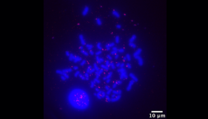
A single GBM39 cell on the CellRaft generated a monoclonal population of cells that formed spheroids in culture and demonstrated a specific genotype as measured by fluorescence in situ hybridization (FISH). 

 

CRT Learn button

team Jessica Hartman
Jessica Hartman, Ph.D.
Senior Director of Product Applications | jessica.hartman@cellmicrosystems.com

Dr. Jessica Hartman has a B.S in Biology from the University of Virginia, a Ph.D. from Duke University in Molecular Cancer Biology and postdoctoral training in Biochemistry and Cancer Biology at Baylor College of Medicine and Duke University, respectively.  She has previously served in Director-level roles, managing bioscience research and development for biotechnology companies. At Cell Microsystems, Dr. Hartman’s role is to lead the development of new and streamlined workflows using the CellRaft Technology and its associated products.

Related Posts

Unlocking the Secrets of Antibiotic Resistance: How Shear Flow Systems Are Revolutionizing the Fight Against Superbugs

In the ongoing battle against antibiotic-resistant bacteria, or “superbugs,” scientists are constantly seeking innovative tools to understand the mechanisms of resistance and develop effective treatments. One such groundbreaking technology is shear flow systems, which have emerged as game-changers in microbiological research. By simulating the dynamic conditions of bacteria’s natural environments, such as the human body or food processing, shear flow systems provide unique insights into bacterial behavior and resistance mechanisms that traditional static methods simply cannot achieve. In this blog post, we’ll explore the benefits of using shear flow systems to combat antibiotic resistance and develop next-generation therapies.

Why Shear Flow Systems Are Essential for Studying Antibiotic Resistance

Antibiotic resistance is a complex phenomenon influenced by a variety of factors, including genetic mutations, horizontal gene transfer, and the formation of biofilms. Traditional laboratory methods often fail to capture the dynamic nature of these processes, leading to incomplete or misleading results. Shear flow systems address this limitation by providing a more realistic environment for bacterial growth and interaction. Here’s how:

1. Mimicking Real-World Conditions

In the human body, bacteria are rarely in a static state. They are constantly exposed to fluid flow, such as blood circulation or urine flow, which influences their behavior and resistance mechanisms. Shear flow systems can replicate these conditions, allowing researchers to study how bacteria respond to antibiotics under realistic physiological conditions. This is critical for understanding how resistance develops and persists in vivo. This benefit was recently highlighted in a webinar presented by Dr. Katharina Richter, a microbiology researcher from the University of Adelaide in Australia. Using a high-throughput BioFlux Shear Flow System, Dr. Richter and her team were able to test 3 different methods of superbug treatments under physiological conditions.

2. Studying Biofilm Formation and Resistance

Biofilms—structured communities of bacteria encased in a protective matrix—are a major contributor to antibiotic resistance. Biofilms are notoriously difficult to treat because they shield bacteria from antibiotics and the immune system. Shear flow systems enable researchers to study biofilm formation in real time, observing how bacteria adhere to surfaces, form microcolonies, and develop resistance under flow conditions. For example, using a BioFlux, Dr. Richter was able to leverage high-resolution imaging to obtain unprecedented insight into the biofilm killing and prevention efficacy of a copper and diethyldithiocarbamate (DDC) combination nanoparticles¹. The group is currently investigating the use of a Cu-DDC infused gel that can be injected into wounds that are at high risk for chronic infection, such as hernia.

From Milliliters to Microliters: How a Shear Flow System Can Reduce Sample Requirements for Experiments

Imagine working with a rare patient-derived cell samples. In a traditional setup, you might need 1 milliliter of sample (equivalent to about 20 drops) to run an experiment. With a microfluidic system, you could potentially get the same results with just 10 microliters (about one-half of a single drop). This efficiency opens up entirely new research possibilities, especially in fields where sample availability is a limiting factor.

Whether due to rarity, cost, or difficulty in obtaining samples, the ability to conduct meaningful experiments with limited sample volumes has long been a goal in biological and chemical research. In this quest for more efficient and precise laboratory techniques, microfluidic shear flow systems have emerged as game-changing tools. These systems are not only reducing the amount of sample required for experiments but are also proving their versatility across a wide range of sample types.

Sample volume

Traditional experimental setups, including static and non-microfluidic shear flow, often demand significant amounts of material, which can be both costly and impractical. This is especially true when working with precious or limited samples such as rare biological specimens, expensive chemicals, or newly synthesized compounds. Enter microfluidic shear flow systems – an elegant solution that’s revolutionizing how investigators approach assays. These miniaturized platforms are proving invaluable across multiple disciplines by dramatically reducing sample requirements from milliliters down to microliters, while maintaining experimental integrity. This up to 1000-fold reduction in sample volume is particularly crucial when working with substances, such as patient-derived cells or novel pharmaceuticals. By significantly reducing the volume of reagents and samples needed, these systems cut costs and make experiments more sustainable.

Sample versatility

Equally as impressive as their sample and reagent reduction ability, is the versatility of microfluidic shear flow systems to adapt to different types of samples.

  • Human Samples: Researchers can study cellular behavior under controlled shear stress, mimicking physiological conditions such as blood or saliva flow.
  • Nanomaterials: The precise fluid dynamics in these systems allow for the uniform dispersion and manipulation of nanoparticles, enabling high-resolution studies in materials science.
  • Microbes: Continuous fluid flow more closely represents the natural environments of many microbes, such as plumbing, food processing, and chronic wounds.

The ability to work with multiple sample types in the same system allows researchers to explore different experimental conditions without the need for multiple specialized setups. By precisely controlling channel geometries and flow rates, researchers can subject different sample types to well-defined shear conditions, enabling detailed studies of cellular mechanics, protein interactions, and material properties.

Microfluidic…
Single Cells: Lab Tune Sung to the Rhythm of "Jingle Bells"

By Jessica Hartman, Ph.D.

Toiling in the lab

With a deadline on its way

To the scope I go

On a Saturday

I don’t see a clone

I’ve looked for one that’s right

What misery these data bring,

I’ll have to look all night,

Oh! Single cells, single cells, dilutions aren’t the way

Oh, I wish I had an AIR to pick a clone, to-da-ay!

Single cells, single cells, I need a better way

Use a Raft to grow a clone or be sad this holiday!

Use a Raft to grow a clone or be sad this holiday!

Now it’s getting late

My spirits are so low

My PI will hate

If my cells don’t grow

My eyesight’s getting dim

A clone I cannot see

My chances are so slim

I need CellRaft Cytometry

Oh! Single cells, single cells, sorters aren’t the way

Oh, I wish I had an AIR to pick a clone, to-da-ay!

Single cells, single cells, I need a better way

Use a Raft to grow a clone or be sad this holiday!

Use a Raft to grow a clone or be sad this holiday!

Oh! Single cells, single cells, dispensers aren’t the way

Oh, I wish I had an AIR to pick a clone, to-da-ay!

Single cells, single cells, I need a better way

Use a Raft to grow a clone or be sad this holiday!

Use a Raft to grow a clone or be sad this holiday!


 “Single Cell” vocalist: Virginia Laurie

Jessica Hartman, Ph.D.Senior Director of Product Applications | jessica.hartman@cellmicrosystems.comDr. Jessica Hartman has a B.S in Biology from the University…