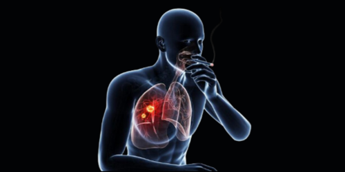
According to the Centers for Disease Control and Prevention, “Tobacco use is the leading cause of preventable disease, disability, and death in the United States. An estimated 28.3 million U.S. adults smoke cigarettes, and about 2.8 million U.S. middle and high school students use at least one tobacco product, including e-cigarettes. Each year, nearly half a million Americans die prematurely of smoking or exposure to secondhand smoke. Another 16 million live with a serious illness caused by smoking” (1).
Smoking tobacco often causes multisystemic diseases affecting various tissues and organs. In particular, epithelial and endothelial cells that line major organs and blood vessels with the structure they need to perform vital functions in the body are targets of nicotine. Research efforts to understand the impact of smoking on the body are hampered by the lack of biologically relevant models and available technologies that can evaluate these complex interactions. As more scientifically relevant tools are developed, scientists can simplify multisystemic diseases into functional models to give researchers better insight into potential treatment options.
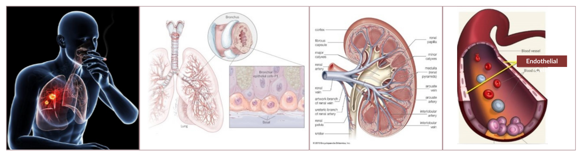 Figure 1. Smoking affects major organs of the body. Bronchial epithelial cells line the structure of the bronchus located within the lungs. HEK293 cells are embryonic kidney cells that would eventually grow to become part of the kidney. Endothelial cells line arteries within the heart where blood cells and monocytes flow through them (3, 4, 8).
Multisystemic diseases give researchers lots of questions to unravel. Importantly, researchers cannot understand all the complex mechanisms that are involved in these diseases with one tool. Using a combination of advanced technology, researchers were able to shed light on some of the questions related to smoking tobacco products.
Bronchial epithelial cells are located within the lungs, providing a protective barrier for airway function. When smoke enters the lungs, bronchial cells are often a target for many carcinogens found within tobacco smoke, causing inflammation within the tissue that can lead to DNA damage, increasing the risk of developing lung cancer (7).
Researchers focused on identifying mutations in DNA caused by smoking tobacco products and compared the degree of mutations with age as well as non-smokers (5). Cells from patients’ lungs were sampled and allowed to settle into microwells of a CellRaft® Array. The CellRaft ArrayConsumables containing thousands of microwells (containing CellRafts) for spatial segregation, imaging, and isolation of single cells, monoclonal colonies, or 3D cultures. More provided special segregation within microwells where single cells could be identified, isolated, and transferred into a PCR tube for genome analysis using the CellRaftA microscale polystyrene growth surface within an array used to spatially segregate cells/colonies while maintaining shared media access, enabling imaging over time and targeted isolation. More AIR® System.
Researchers uncovered that the frequency of mutations in DNA correlating to smoking tobacco products leveled off with people who smoked 23 packs per year.

(Left) CellRaft ArrayConsumables containing thousands of microwells (containing CellRafts) for spatial segregation, imaging, and isolation of single cells, monoclonal colonies, or 3D cultures. More before and after isolation. The needle was used to dislodge the CellRaftA microscale polystyrene growth surface within an array used to spatially segregate cells/colonies while maintaining shared media access, enabling imaging over time and targeted isolation. More containing the cell from the CellRaft ArrayConsumables containing thousands of microwells (containing CellRafts) for spatial segregation, imaging, and isolation of single cells, monoclonal colonies, or 3D cultures. More. A 0.2-mL PCR tube with 2.5 µL PBS containing one CellRaftA microscale polystyrene growth surface within an array used to spatially segregate cells/colonies while maintaining shared media access, enabling imaging over time and targeted isolation. More was collected by the CellRaft AIR SystemAn all-in-one platform for imaging, identifying, and isolating viable single cells, colonies, or organoids using CellRaft Arrays and software-guided selection with CellRaft Cytometry. More (magnified). The small brown dot at the bottom of the PCR tube is the raft (arrow). Using this system it is easy to visually check if more than one cell was captured in the same tube. (Right) Schematic representation of the isolation, processing, and analysis of PBBCs from human lung.
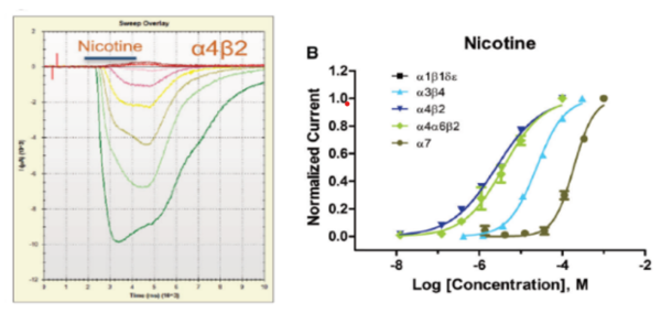
Nicotine is the main psychoactive ingredient in tobacco products, acting as a stimulant within the body. Nicotine interacts with acetylcholine receptors in the brain (nAChRs). Once activated, these receptors conduct positively charged ions like sodium and calcium across the cell membrane leading to cell excitation. Using stably transfected HEK293 cells, researchers can study the effect of nicotine on different transfected receptors. Recently, nicotine concentration-dependent responses were assessed against multiple targets stably expressed in HEK293 cells using an IonFlux Mercury automated patch clamp system (9). When the compounds reached the cells and activated the receptors, currents were elicited and measured to determine the EC50 (half maximal effective concentration) of these receptors. As expected nicotine had no effect on the primary nAChR from the neuromuscular junction (α1β1δε), while having effects on other receptors with the EC50 reported as follows: α3β4 (23.9 µM), α4β2 (2.54 µM), α4α6β2 (3.79 µM), and α7 (188.4 µM).
In addition to traditional tobacco smoke produced from cigarettes, a new generation of smoking has become widespread with the use of electronic cigarettes that deliver nicotine vapor into the lungs. Smoking plays a major role in the pathogenesis and progression of atherosclerosis, as it damages arteries, allowing fatty deposits to form, which can develop into plaques. These plaques contribute to vessel hardening and reduce lumen size, which increases the chances of developing blockages. Monocytes are released into the blood to help protect the body from pathogens. When smoke enters the blood vessels, it affects the endothelial surface, increasing monocyte adherence, which contributes to the formation of atherosclerosis causing plaque.
In this experiment, human aortic endothelial cells (HAECs) represented the endothelial surface of cardiac vessels and THP-1 (human acute monocytic leukemia) cells were the monocytes protecting the body from invaders like smoke (6). HAECs were grown in a monolayer within the microfluidic channels to form “vessel-like” surfaces while THP-1 cells flowed in and adhered to HAECs in response to treatment of whole smoke conditioned media (WSCM) or electronic cigarette vapor conditioned media (eVCM).
The use of the BioFlux microfluidic flow plates allowed THP-1 adhesion to endothelial cells to be evaluated under physiological shear flow, emulating in vivo conditions. As THP-1 cell adhesion was measured to evaluate that as the concentration of WSCM increased, monocyte adhesion compared to the control for either condition showed no increases. They discovered that the increase in monocyte adhesion in the WSCM was linked to a regulator for cell adhesion expressed from endothelial cells.
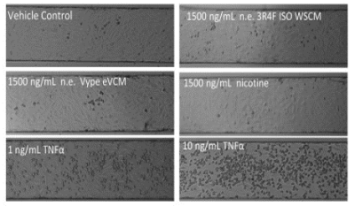 Figure 4. Impact of cigarette versus electronic cigarette aerosol conditioned media on aortic endothelial cells in a microfluidic cardiovascular model (6).
In these examples, researchers had diverse biological questions related to the effects of smoking tobacco products. Each group shared a similar research goal while providing different discoveries with advanced technologies. The tools used in these publications can be combined to add additional insight to many angles of research. The expansion into scientific discoveries that are enabled using the CellRaft AIR SystemAn all-in-one platform for imaging, identifying, and isolating viable single cells, colonies, or organoids using CellRaft Arrays and software-guided selection with CellRaft Cytometry. More, IonFlux Mercury, and BioFlux provide researchers with appropriate resources to uncover their own discoveries in their field of study. Whether you are developing models or analyzing cells to answer clues about specific problems, the right tool can lead to faster solutions accelerating your research into the next generation.
“A good tool improves the way you work. A great tool improves the way you think.” – Jeff Duntemann

If you are ready to uncover scientific discoveries related to complicated processes, check out how our novel technology can accelerate your research: CellRaft AIR, IonFlux Mercury, and BioFlux
References

Lexi Land contributes to developing scientific workflows that are compatible with the CellRaft® AIR System. Her focus at Cell Microsystems involves stem cell research, 3D organoid work, as well as adherent and suspension cell culture. Lexi received a Bachelor of Science in Biological Sciences from North Carolina State University with a focus in molecular, cellular, and developmental biology.