Blog

Go with the Flow: Shear Stress Enhances Physiological Relevance

In vivo, most cell types exist surrounded by moving fluid that influences their phenotype and function. The flow created from moving fluids creates shear stress, which can influence cytokine production, polarization, transcription factor expression, organization, and morphology of many cell types (Tzima, E., 2005, Shi, X., 2016, Chen, C., 2014). However, despite these facts, many biological investigations are conducted in vitro. Despite the convenience of in vitro cell cultures for modeling physiological processes, in vitro cultures are critically limited by the static nature of the culture system. This limits the translation of in vitro results to in vivo systems. Therefore, the ability to accurately mimic in vivo environments using physiological shear flow is critical to understanding the true biological responses of cells. Here, we highlight several flow-dependent biological processes that cannot easily be studied in traditional cell culture dishes, as well as a simple solution to investigate these cells under shear flow.


Figure 1. Shear stress on the vascular wall
shear stress in vessel diagram
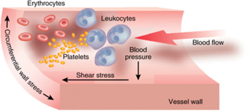
Figure 1. Diagram of the major hemodynamic stress forces on the vascular wall. The forces acting on a cell can be described in terms of pressure or stress, which is defined as force per unit area, for example, pascal (Pa). In vascular biology, the stress is ‘shear’ when the force is applied parallel to the area and ‘normal’ when the force is perpendicular to the area. Major hemodynamic stress forces include fluid shear stress, circumferential stress, and blood pressure. (Li, L., Terry, 2008)


Atherosclerosis

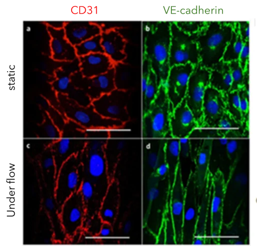
Atherosclerosis can lead to restricted blood flow, causing a range of complications. The restriction in flow is due to a combination of factors, including reduced nitric oxide (NO) bioavailability, which impairs vessel dilation, and plaque accumulation, which reduces the size of the vessel, thereby limiting blood flow. Flow of different levels, high to low, can be utilized to simulate different parts of this disease to elucidate the mechanisms of atherosclerosis. For example, shear flow not only impacts endothelial cell alignment (Figure 2) but also influences signaling pathways and gene expression within endothelial cells. Furthermore, leukocyte adhesion to the endothelium and transmigration into the arterial intima, important steps in the development and progression of atherosclerosis, are impacted by shear flow. Therefore, accurate in vitro investigation of atherosclerosis mechanisms in a simulated in vivo environment can speed discovery and development of atherosclerosis treatments and preventions.


Figure 2. Impact of shear flow on endothelial cells

cd31 ve caherin with and without shear flow new axis labels
Figure 2. Endothelial cell characterization and overview of cell distribution in microfluidic channels both under static and flow conditions. Endothelial cell characterization by expression of CD31 and VE-cadherin. (a–b). Representative images for CD31 and VE-cadherin expression under static conditions; (c–d) representative images for CD31 and VE-cadherin expression under flow conditions. Scale bars represent 50 µm. In cells cultured under static conditions, CD31 and VE-cadherin were expressed in arbitrary patterns, whereas CD31 and VE-cadherin were aligned with the direction of the pulsatile flow when the cells were cultured for 2 days at 10 dyn/cm2. This indicates that the expression of these endothelial cell markers is affected by shear stress-dependent mechanotransduction (Sfriso, R.,2018).


Blood Clotting

Platelets are critical for the return to homeostasis following physical wounding, as they bind to damaged blood vessels to prevent excessive bleeding. Additionally, platelets play a major role in multiple diseases, including sickle cell disease, atherosclerosis, and hemophilia. Dysregulated platelet function can lead to poor clotting and subsequent bleeding or to the formation of thrombi, which can occlude blood flow to tissue. Although aggregometry is typically used to study the mechanisms of platelet function, this method is low-throughput, requires relatively large amounts of blood, and perhaps most importantly, is not performed under flow conditions. These critical limitations reduce the relevance of many platelet investigations because platelets can be activated by high shear stress, such as injuries to capillaries or turbulent flow near atherosclerotic plaques. The ability to investigate and image platelet interactions and thrombus formation under physiological and pathological shear flow, aids the understanding of the mechanisms that contribute to platelet function.


Video 1.  Thrombin-loaded injury-site-targeted lipid nanoparticles

Video 1. t-TLNPs (thrombin-loaded injury-site-targeted lipid nanoparticles) enhance fibrin generation under simulated vascular flow environment imaged in real-time using BioFlux microfluidic setup. Human plasma containing fluorescently labeled platelets and fibrinogen (by Calcein and AlexaFluor647, respectively) (Girish, A., 2022).


Cancer cells

Cancer cell chemotaxis and transmigration contribute to metastasis and other oncological processes in the vascular system. Examining migration over time under shear flow can help screen compounds that inhibit cancer cell invasion, leading to better treatment options. Furthermore, tumors are known to alter the microenvironments that surround them. This can promote immunotolerance, encourage tumor angiogenesis, and negatively impact nearby tissue. In vivo examination of these responses is difficult due to the heterogeneity of the cells in the tumor’s environment. Stepwise mechanistic examination of tumors and their impact on the surrounding microenvironment is much more feasible in a controlled modular system that simulates in vivo conditions.


Figure 3. Shear stress by vessel
range of shear stress in the body1
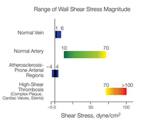
Figure 3. Shear stress values in different human vessel types. Values derived from studies by AM Malek et al., (JAMA, 1999).


CAR-T cells

Immunotherapies like CAR-T and TCR require interactions between multiple cell types and target cells and tissues. Real-time cell adhesion, transmigration, binding strength, and other aspects of T cell therapies can be assessed under physiological flow as compounds are introduced. In addition, this type of tightly controlled assay can enhance CAR-T therapy by ensuring that engineered cells have the desired functional properties before delivering treatment to patients.


Bacterial and Fungal Biofilms

Bacteria and fungi often form biofilms, which are complex communities of microbes bound by an extracellular matrix. Biofilms are beneficial to microbes for various reasons, including adhesion, nutrient cycling, exchange of genetic material, and division of metabolic labor amongst different subpopulations. However, the development of biofilms is often detrimental to humans, such as in the case of plaque accumulation on teeth and medical device infection. Although biofilms can be investigated in traditional cultures, the static nature of the culture can lead to nutrient depletion and metabolite accumulation, which does not reflect natural systems where biofilms modulate their response to environmental changes. Therefore, efficient development of antibiotic and antimicrobial drugs should be done under shear flow conditions.      


Figure 4. Confocal Laser Scanning Microscopy of viable P338-adsorbed and unadsorbed biofilms
biofilm stirpe et al
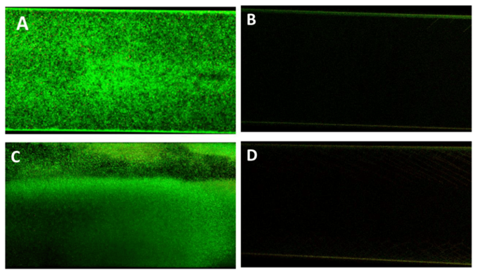
Figure 4. Antifouling activity of P388 in dynamic conditions. Representative CLSM images of Ec5FSL (A,B) and Ec9FSL (C,D) biofilms formed after 15 h under a shear flow of 0.5 dyn/cm2 (0.05 Pa) on unadsorbed (A,C) and P388-adsorbed (B,D) microfluidic channel (Stirpe et al. 2020).

Conclusions

Accurate investigation of processes that are impacted by flow in the body requires similar flow in vitro to examine relevant responses. Instruments such as BioFlux can create shear stress at specific intensities that can be adjusted to mimic the physiological or pathological shear stress that cells of interest experience in vivo. A key component of the BioFlux system is the ability to provide real-time imaging, enabling a clearer understanding of the interactions between cells or the effects of compounds during drug discovery and development investigations. Adding shear stress to cells in the form of flow can provide many benefits to in vitro experiments that induce physiological responses similar to those that occur in vivo.


References

  1. Chen, C., & Khismatullin, D. B. (2014). Lipopolysaccharide induces the interactions of breast cancer and endothelial cells via activated monocytes. Cancer Letters, 345(1), 75–84. https://doi.org/10.1016/j.canlet.2013.11.022
  2. Girish, A., Jolly, K., Alsaadi, N., de la Fuente, M., Recchione, A., An, R., Disharoon, D., Secunda, Z., Raghunathan, S., Luc, N. F., Desai, C., Knauss, E., Han, X., Hu, K., Wang, H., Sekhon, U. D. S., Rohner, N., Gurkan, U. A., Nieman, M., Neal, M. D., … Sen Gupta, A. (2022). Platelet-Inspired Intravenous Nanomedicine for Injury-Targeted Direct Delivery of Thrombin to Augment Hemostasis in Coagulopathies. ACS nano, 16(10), 16292–16313. https://doi.org/10.1021/acsnano.2c05306
  3. Li, L., Terry, C. M., Shiu, Y. T., & Cheung, A. K. (2008). Neointimal hyperplasia associated with synthetic hemodialysis grafts. Kidney international, 74(10), 1247–1261. https://doi.org/10.1038/ki.2008.318
  4. Malek AM, Alper SL, Izumo S. Hemodynamic shear stress and its role in atherosclerosis. JAMA. 199 Dec 1; 282(21):2035-42. doi: 10.1001Tjama.282.21.2035.
  5. Sfriso, R., Zhang, S., Bichsel, C. A., Steck, O., Despont, A., Guenat, O. T., & Rieben, R. (2018). 3D artificial round section micro-vessels to investigate endothelial cells under physiological flow conditions. Scientific Reports, 8(1). https://doi.org/10.1038/s41598-018-24273-7
  6. Shi, X., Yang, J., Huang, J., Long, Z., Ruan, Z., Xiao, B., & Xi, X. (2016). Effects of different shear rates on the attachment and detachment of platelet thrombi. Molecular medicine reports, 13(3), 2447–2456. https://doi.org/10.3892/mmr.2016.4825
  7. Tzima, E. et al. A mechanosensory complex that mediates the endothelial cell response to fluid shear stress. Nature 437, 426–431 (2005).
  8. Stirpe M, Brugnoli B, Donelli G, Francolini I, Vuotto C. Poloxamer 338 Affects Cell Adhesion and Biofilm Formation in Escherichia coli. Potential Applications in the Management of Catheter-Associated Urinary Tract Infections. Pathogens. 2020; 9(11):885. https://doi.org/10.3390/pathogens9110885
Lexi Land B.S. 300x300 1
Lexi Land, B.S.
Research Associate II | lland@cellmicrosystems.com

Lexi Land contributes to developing scientific workflows that are compatible with the CellRaft® AIR System. Her focus at Cell Microsystems involves stem cell research, 3D organoid work, as well as adherent and suspension cell culture. Lexi received a Bachelor of Science in Biological Sciences from North Carolina State University with a focus in molecular, cellular, and developmental biology.
Related Posts

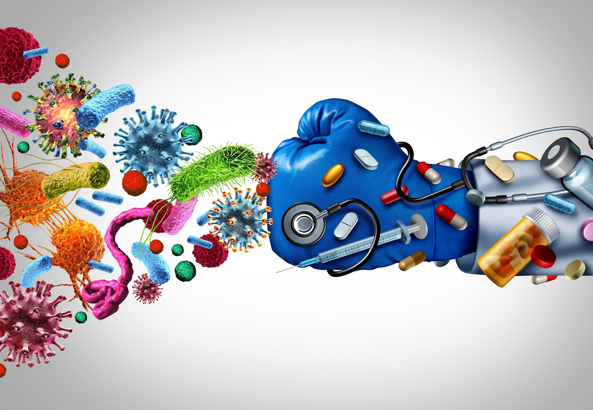
Unlocking the Secrets of Antibiotic Resistance: How Shear Flow Systems Are Revolutionizing the Fight Against Superbugs

In the ongoing battle against antibiotic-resistant bacteria, or “superbugs,” scientists are constantly seeking innovative tools to understand the mechanisms of resistance and develop effective treatments. One such groundbreaking technology is shear flow systems, which have emerged as game-changers in microbiological research. By simulating the dynamic conditions of bacteria’s natural environments, such as the human body or food processing, shear flow systems provide unique insights into bacterial behavior and resistance mechanisms that traditional static methods simply cannot achieve. In this blog post, we’ll explore the benefits of using shear flow systems to combat antibiotic resistance and develop next-generation therapies.

Why Shear Flow Systems Are Essential for Studying Antibiotic Resistance

Antibiotic resistance is a complex phenomenon influenced by a variety of factors, including genetic mutations, horizontal gene transfer, and the formation of biofilms. Traditional laboratory methods often fail to capture the dynamic nature of these processes, leading to incomplete or misleading results. Shear flow systems address this limitation by providing a more realistic environment for bacterial growth and interaction. Here’s how:

1. Mimicking Real-World Conditions

In the human body, bacteria are rarely in a static state. They are constantly exposed to fluid flow, such as blood circulation or urine flow, which influences their behavior and resistance mechanisms. Shear flow systems can replicate these conditions, allowing researchers to study how bacteria respond to antibiotics under realistic physiological conditions. This is critical for understanding how resistance develops and persists in vivo. This benefit was recently highlighted in a webinar presented by Dr. Katharina Richter, a microbiology researcher from the University of Adelaide in Australia. Using a high-throughput BioFlux Shear Flow System, Dr. Richter and her team were able to test 3 different methods of superbug treatments under physiological conditions.

2. Studying Biofilm Formation and Resistance

Biofilms—structured communities of bacteria encased in a protective matrix—are a major contributor to antibiotic resistance. Biofilms are notoriously difficult to treat because they shield bacteria from antibiotics and the immune system. Shear flow systems enable researchers to study biofilm formation in real time, observing how bacteria adhere to surfaces, form microcolonies, and develop resistance under flow conditions. For example, using a BioFlux, Dr. Richter was able to leverage high-resolution imaging to obtain unprecedented insight into the biofilm killing and prevention efficacy of a copper and diethyldithiocarbamate (DDC) combination nanoparticles¹. The group is currently investigating the use of a Cu-DDC infused gel that can be injected into wounds that are at high risk for chronic infection, such as hernia.

From Milliliters to Microliters: How a Shear Flow System Can Reduce Sample Requirements for Experiments

Imagine working with a rare patient-derived cell samples. In a traditional setup, you might need 1 milliliter of sample (equivalent to about 20 drops) to run an experiment. With a microfluidic system, you could potentially get the same results with just 10 microliters (about one-half of a single drop). This efficiency opens up entirely new research possibilities, especially in fields where sample availability is a limiting factor.

Whether due to rarity, cost, or difficulty in obtaining samples, the ability to conduct meaningful experiments with limited sample volumes has long been a goal in biological and chemical research. In this quest for more efficient and precise laboratory techniques, microfluidic shear flow systems have emerged as game-changing tools. These systems are not only reducing the amount of sample required for experiments but are also proving their versatility across a wide range of sample types.

Sample volume

Traditional experimental setups, including static and non-microfluidic shear flow, often demand significant amounts of material, which can be both costly and impractical. This is especially true when working with precious or limited samples such as rare biological specimens, expensive chemicals, or newly synthesized compounds. Enter microfluidic shear flow systems – an elegant solution that’s revolutionizing how investigators approach assays. These miniaturized platforms are proving invaluable across multiple disciplines by dramatically reducing sample requirements from milliliters down to microliters, while maintaining experimental integrity. This up to 1000-fold reduction in sample volume is particularly crucial when working with substances, such as patient-derived cells or novel pharmaceuticals. By significantly reducing the volume of reagents and samples needed, these systems cut costs and make experiments more sustainable.

Sample versatility

Equally as impressive as their sample and reagent reduction ability, is the versatility of microfluidic shear flow systems to adapt to different types of samples.

  • Human Samples: Researchers can study cellular behavior under controlled shear stress, mimicking physiological conditions such as blood or saliva flow.
  • Nanomaterials: The precise fluid dynamics in these systems allow for the uniform dispersion and manipulation of nanoparticles, enabling high-resolution studies in materials science.
  • Microbes: Continuous fluid flow more closely represents the natural environments of many microbes, such as plumbing, food processing, and chronic wounds.

The ability to work with multiple sample types in the same system allows researchers to explore different experimental conditions without the need for multiple specialized setups. By precisely controlling channel geometries and flow rates, researchers can subject different sample types to well-defined shear conditions, enabling detailed studies of cellular mechanics, protein interactions, and material properties.

Microfluidic…
Single Cells: Lab Tune Sung to the Rhythm of "Jingle Bells"

By Jessica Hartman, Ph.D.

Toiling in the lab

With a deadline on its way

To the scope I go

On a Saturday

I don’t see a clone

I’ve looked for one that’s right

What misery these data bring,

I’ll have to look all night,

Oh! Single cells, single cells, dilutions aren’t the way

Oh, I wish I had an AIR to pick a clone, to-da-ay!

Single cells, single cells, I need a better way

Use a Raft to grow a clone or be sad this holiday!

Use a Raft to grow a clone or be sad this holiday!

Now it’s getting late

My spirits are so low

My PI will hate

If my cells don’t grow

My eyesight’s getting dim

A clone I cannot see

My chances are so slim

I need CellRaft Cytometry

Oh! Single cells, single cells, sorters aren’t the way

Oh, I wish I had an AIR to pick a clone, to-da-ay!

Single cells, single cells, I need a better way

Use a Raft to grow a clone or be sad this holiday!

Use a Raft to grow a clone or be sad this holiday!

Oh! Single cells, single cells, dispensers aren’t the way

Oh, I wish I had an AIR to pick a clone, to-da-ay!

Single cells, single cells, I need a better way

Use a Raft to grow a clone or be sad this holiday!

Use a Raft to grow a clone or be sad this holiday!


 “Single Cell” vocalist: Virginia Laurie

Jessica Hartman, Ph.D.Senior Director of Product Applications | jessica.hartman@cellmicrosystems.comDr. Jessica Hartman has a B.S in Biology from the University…