Single cell workflows are hot topics in scientific research today. The ability to turn a single cell into a viable monoclonal colony is essential for cell line development, recombinant protein, and antibody production, and induced pluripotent stem cell engineering. Such monoclonal cell lines are the foundational building blocks enabling cutting-edge disease modeling, drug screening, and personalized medicine.
Unfortunately, one of the most challenging steps in these workflows is one of the first steps – single cell isolation and monoclonal expansion. To generate single cell colonies, researchers often utilize limiting dilutionA statistical cloning approach where cells are diluted to low density to “hopefully” seed one cell per well; commonly used but can be low-efficiency for difficult-to-clone lines. More. However, this technique is tremendously time-consuming and has very low efficiency. Other techniques such as fluorescence activated cell sorting (FACS) and single cell droplet dispensing are common, but the fluidics and cell manipulations required often negatively impact cell viability (Cell Line Development Raftnote (cellmicrosystems.com). Those cells that are successfully seeded as single cells, typically in 96-well or 384-well plates, then must overcome the hurdle of growing and forming viable clones.
Cell growth and division depend on a number of elements, including growth factors and mitogens that regulate the cell cycle to promote cell division. Under normal physiological conditions in the body, cells grow together and communicate with each other to function as a unit and enhance growth and survival. This is recapitulated in typical tissue culture conditions where cells are grown together in flasks, and they secrete growth factors which act in a paracrine manner to promote cell growth amongst the entire population. Problems in cell growth and proliferation can occur when there are too many cells within a dish, depleting nutrients in the media and resulting in cell cycle arrest. On the other hand, when cells are plated as single cells or at low density, they no longer receive growth factor signals .
To promote cell growth during single cell workflows, mitogens, such as Platelet-Derived Growth Factor, are provided to cells through the addition of serum to the medium. Growth factors can also be supplemented to the medium to stimulate cell growth (Extracellular Control of Cell Division, Cell Growth, and Apoptosis – Molecular Biology of the Cell – NCBI Bookshelf (nih.gov)). However, serums can vary from batch to batch and many manufacturing processes require animal-free products (Media and Supplements in Cell Culture (sigmaaldrich.com)). Furthermore, individual growth factors, such as basic Fibroblast Growth Factor (bFGF) and Epidermal Growth Factor (EGF) cost hundreds of dollars for small weights, adding additional costs to tissue culture workflows.
To overcome the challenge of single cell viability and outgrowth, researchers often choose to plate cells in conditioned media – media obtained from confluent cell culture dishes that contain all the extracellular growth factors secreted by the cells to promote the growth of the single cell colony. However, this process is time-consuming and can be variable from batch to batch. Specialized medium and supplements can also be purchased, but these are expensive and the cloning efficiency of single cells plated with these supplements is still only around ~30% (InstiGRO CHO and HEK | Animal-Free Cell Culture Supplements (salscientific.com) )
Fundamentally, any technology that is utilized for single cell cloning that deposits a single cell into a well of a tissue culture dish will contend with the hurdle of a single cell trying to divide in a way that is not biologically familiar. Ideally, to enable high-efficiency monoclonal cell line development, cells need to be cultured in a flask-like bulk culture environment that also provides spatial single cell segregation.
A novel solution that enables this possibility is the CellRaft® Technology from Cell Microsystems. Cells are seeded onto CellRaftA microscale polystyrene growth surface within an array used to spatially segregate cells/colonies while maintaining shared media access, enabling imaging over time and targeted isolation. More arrays, which are a flask-like tissue culture dish in which seeded cells share culture media, while also being spatially segregated within elastomeric microwells. Each microwell contains a ferric, polystyrene tissue culture-coated growth surface, called a CellRaftA microscale polystyrene growth surface within an array used to spatially segregate cells/colonies while maintaining shared media access, enabling imaging over time and targeted isolation. More, and each CellRaft arrayConsumables containing thousands of microwells (containing CellRafts) for spatial segregation, imaging, and isolation of single cells, monoclonal colonies, or 3D cultures. More contains thousands to tens of thousands of individual microwells. Low-density cell suspensions can be seeded onto the array, and the cells settle by gravity and adhere to the CellRafts. Thus, the attached cells can grow on the CellRaftA microscale polystyrene growth surface within an array used to spatially segregate cells/colonies while maintaining shared media access, enabling imaging over time and targeted isolation. More and maintain spatial single cell integrity, while also communicating through contiguous medium. This unique feature leads to dramatically increased cell viability and single cell clone formation. Importantly, during the growth phase, the CellRaft arrayConsumables containing thousands of microwells (containing CellRafts) for spatial segregation, imaging, and isolation of single cells, monoclonal colonies, or 3D cultures. More can be easily imaged using the CellRaft AIR SystemAn all-in-one platform for imaging, identifying, and isolating viable single cells, colonies, or organoids using CellRaft Arrays and software-guided selection with CellRaft Cytometry. More, and CellRafts containing single cells can be identified and monitored as they undergo cell division. CellRafts containing clones of interest can be easily retrieved using the CellRaft AIR systemAn all-in-one platform for imaging, identifying, and isolating viable single cells, colonies, or organoids using CellRaft Arrays and software-guided selection with CellRaft Cytometry. More for further expansion in 96-well collection plates. Because the clones are being isolated for downstream growth as small colonies at this stage rather than as single cells, they have already overcome the hurdle of growing out of the single cell stage and the success rate is dramatically higher.
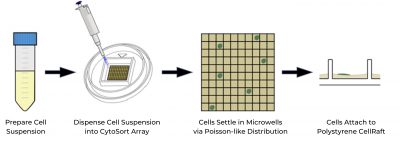
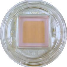
One example of how the CellRaft ArrayConsumables containing thousands of microwells (containing CellRafts) for spatial segregation, imaging, and isolation of single cells, monoclonal colonies, or 3D cultures. More improves single cell viability and clonal outgrowth comes from a case study where a researcher reached out to us for help generating a monoclonal cell line from an edited polyclonal population. Previous workflows in their lab involved producing conditioned media and then performing FACS or limiting dilutionA statistical cloning approach where cells are diluted to low density to “hopefully” seed one cell per well; commonly used but can be low-efficiency for difficult-to-clone lines. More into the conditioned media in the hope of isolating a single clone. Within one week, we were able to seed the sensitive cells onto the array, identify single cells, and isolate over 100 single cell-derived colonies. As the single cells were able to share media on the array just like they would in a flask, there was no need for conditioned media in our experiment, saving the researcher time and resources.
This case study highlights the importance of providing paracrine cell-cell communication in order to promote single cell growth and viability. In this example, the same cell population that failed to produce even a single viable edited colony after single-cell culture was able to generate hundreds of monoclonal colonies on the CellRaft arrayConsumables containing thousands of microwells (containing CellRafts) for spatial segregation, imaging, and isolation of single cells, monoclonal colonies, or 3D cultures. More, simply by changing the culture environment and avoiding the pitfalls of single-cell culture.


Dr. Schoepke received post-doctoral training in translational breast cancer research at Baylor College of Medicine, obtained a Ph.D. in Pharmacology and Physiology from Saint Louis University, and holds a Bachelor of Science in Molecular and Cellular Biology with a minor in Chemistry from the University of Illinois at Urbana-Champaign. Her background is in Nuclear Receptor pharmacology, testing novel drugs in 2D and 3D cell-based assays of cancer and in vitro models of metabolic disease and exercise. Dr. Schoepke is currently a Field Application Scientist at Cell Microsystems who focuses on demonstrating and training new customers on the CellRaft® AIR System as well as troubleshooting novel single cell workflows.