The World Health Organization stated that as of 2020, nearly 8 million women alive were diagnosed with breast cancer in the last 5 years, making it the world’s most prevalent cancer (Retrieved from WHO). Globally, 1 in 8 women will be affected at some point during their lives with breast cancer and must undergo surgery or radiation treatment options that still include invasive and damaging methods developed over 50 years ago.
Aiding in the development of new treatment modalities that are noninvasive with fewer side effects leads to the evolution of breast cancer cell models. These new methods shine a light of hope on a very dark past for individuals who have undergone radiation treatment only to fail treatment or relapse. While these treatment options offer substantial promise, they often take nearly a decade or longer to complete all phases of clinical trials before full FDA approval.
To begin the process for a clinical trial, cancer models are created from cell lines to understand the in vitro and in vivo effects of the drugs before being tested in people. Only after being cleared from having any toxic effects will the drug be moved further into clinical research with humans. After that, the FDA will review and determine a drug to be safe and effective against its intended use and offer recommendation for approval.
One aggressive subtype of breast cancer is HER2 positive disease. HER2 is a human epidermal growth factor receptor that normally acts to maintain cell functions for growth and development of breast cells. When this gene is overexpressed, it leads to aberrant expression of the receptor and tumor formation. Approximately 10-20% of breast cancers are traced back to HER2 receptors (Figure 1).
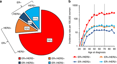
Figure 1: Distribution of the breast cancer subtypes by ER/HER2 status and their age-specific incidence in Scotland for 2009–2016 (N = 31,099). a Shows a pie chart and b shows age-specific incidence on the log scale by subtype. b Data are for 31,099 breast cancer cases with ER/HER2 missing status imputed for analysis. Dotted lines in the graph denote ages 50–70 years, the age group invited for screening in Scotland every 3 years. (Retrieved from Nature, 2020)
Cellular models provide consistency and reliability for in vitro and in vivo testing pre-clinical trials. To ensure stability and genetic reproducibility, it is important to derive a cellular model from a monoclonal population. Without uniform models, results can be unreliable and discredit potential treatment methods that may be successful and provide improved outcomes for cancer patients.
Despite the importance of such models, the actual development of cell lines in the lab is a time-consuming hurdle that necessitates substantial effort. These efforts are often met with failure due to technical challenges and limitations.
To study aggressive HER2+ breast cancer, a cell line was engineered to overexpress HER2 and blue fluorescent protein (EBFP2). The researchers generating this line leveraged a traditional method for deriving a stable monoclonal cell population, fluorescence activated cell sorting (FACS), which involves using laser excitation and fluidics to recover single cells. However, FACS often results in an unstable cell population with varying degrees of expression of the gene of interest. This inevitability dilutes or ultimately obscures downstream analysis, making reliability and reproducibility impossible. After two separate rounds of FACS to enrich the double positive population (HER2+/EBFP2+), the cells nonetheless remained heterogeneous. This caused an inherent competition for survival in the population and the genetic edits became diluted and unstable.
Realizing the cellular population was quickly fading and the correct edits were being lost in the bulk population, a monoclonal and stable cell line needed to be generated quickly. By seeding the population on a CellRaft ArrayConsumables containing thousands of microwells (containing CellRafts) for spatial segregation, imaging, and isolation of single cells, monoclonal colonies, or 3D cultures. More, clonality was able to be tracked and traced from day 0, as well as expression of the EBFP2 marker. This ensured chain of custody in the development of each cell colony (Figure 2). In addition to clonality, it was also necessary to identify cells expressing high levels of HER2. Using a live stain for HER2 on the CellRaft AIR SystemAn all-in-one platform for imaging, identifying, and isolating viable single cells, colonies, or organoids using CellRaft Arrays and software-guided selection with CellRaft Cytometry. More, monoclonal populations that were HER2 positive as well as EBFP2 positive were discovered. When interrogating the population of cells on the CellRaft ArrayConsumables containing thousands of microwells (containing CellRafts) for spatial segregation, imaging, and isolation of single cells, monoclonal colonies, or 3D cultures. More, it was clear that the actual population of double expressing cells was extremely rare compared to the data generated from FACS. Approximately 1.5% of the population of cells retained the correct gene edits compared to 89% reported from sorting. The AIR System was able to isolate populations that met all criteria, and they were expanded for cryopreservation.
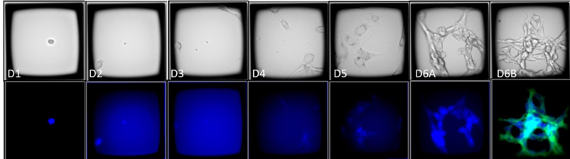
Figure 2: Time course images of a CellRaftA microscale polystyrene growth surface within an array used to spatially segregate cells/colonies while maintaining shared media access, enabling imaging over time and targeted isolation. More containing an EBFP2+/HER2+ clone. The CellRaftA microscale polystyrene growth surface within an array used to spatially segregate cells/colonies while maintaining shared media access, enabling imaging over time and targeted isolation. More arrays were imaged in brightfield and fluorescence 4 hours post-seeding and every 24 hours until colony formation. On day 6, the array was stained with Anti-HER2-FITC to visualize HER2 expression (green). 10X Magnification.
This workflow enabled thousands of cells to be screened using a single consumable to identify the rare double positive clones in a matter of weeks, not months. In total, the time from cell seeding to clonal outgrowth was only 16 days, and viable cell banks of the verified clones were cryopreserved in 27 days. Thus, the CellRaftA microscale polystyrene growth surface within an array used to spatially segregate cells/colonies while maintaining shared media access, enabling imaging over time and targeted isolation. More AIR Technology enables successful monoclonal cell line development for even rare or challenging cells faster and more efficiently than traditional methods.
For an in-depth look at this case study, download: Identification of a rare double positive clone.
Learn more about CellRaftA microscale polystyrene growth surface within an array used to spatially segregate cells/colonies while maintaining shared media access, enabling imaging over time and targeted isolation. More Technology.

Lexi Land contributes to developing scientific workflows that are compatible with the CellRaft® AIR System. Her focus at Cell Microsystems involves stem cell research, 3D organoid work, as well as adherent and suspension cell culture. Lexi received a Bachelor of Science in Biological Sciences from North Carolina State University with a focus in molecular, cellular, and developmental biology.