Blog

Advanced Therapy Medicinal Products – FDA recommended pathways for manufacturers and need for single cell cloning


FDA Regulatory pathways

The U.S. Food and Drug Administration (FDA) established the Office of Tissues and Advanced Therapies (OTAT) to oversee the development and regulation of advanced therapy medicinal products (ATMPs). (ATMPs are a class of medicinal products that include gene therapies, cell therapies, and tissue-engineered products.) OTAT works with other FDA offices and centers to ensure that ATMPs are developed and regulated in a way that is consistent with the FDA’s mission to protect public health.


Regulatory Pathways

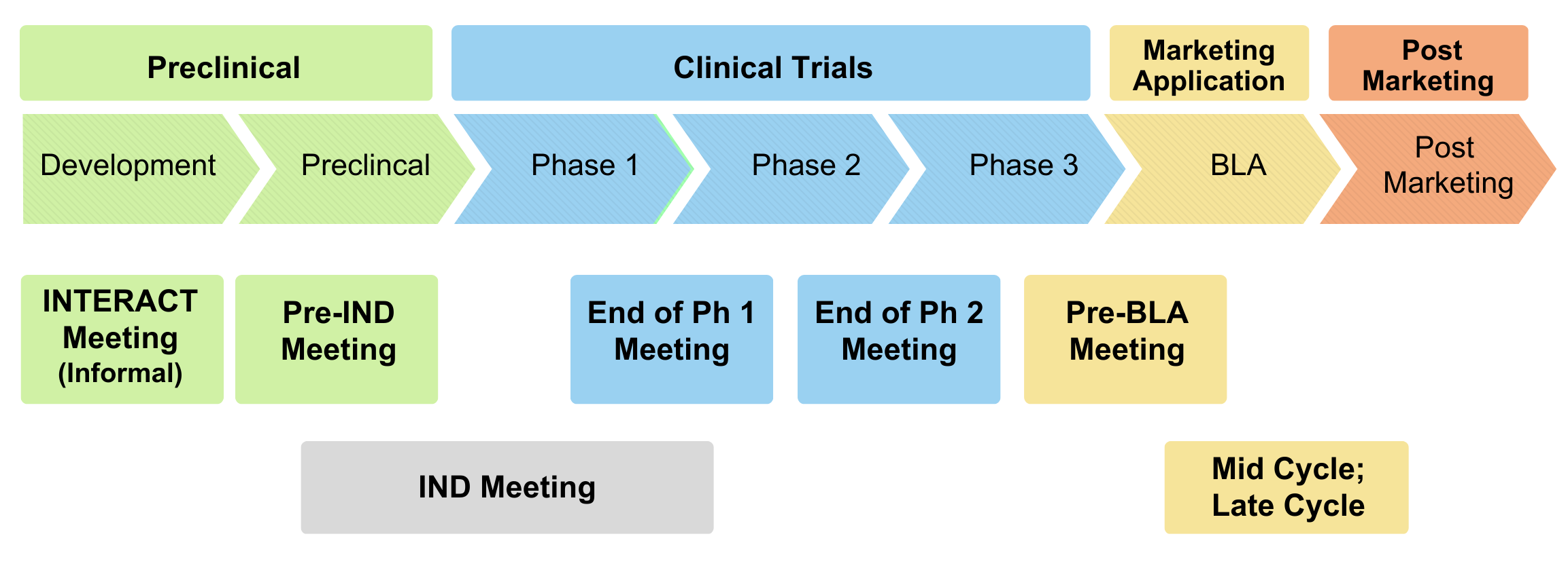
The FDA has established several regulatory pathways for ATMPs, including:

Investigational New Drug (IND) pathway: This pathway is for ATMPs that are in the early stages of development and are not yet approved for marketing. Sponsors of INDs must submit an IND application to the FDA, which includes data on the safety and effectiveness of the ATMP.

Biologics License Application (BLA) pathway: This pathway is for ATMPs that have completed clinical testing and are ready for marketing. Sponsors of BLAs must submit a BLA to the FDA, which includes data on the safety, effectiveness, and manufacturing of the ATMP.

Humanitarian Device Exemption (HDE) pathway: This pathway is for ATMPs that are intended to treat or diagnose rare diseases or conditions. Sponsors of HDE applications must submit an HDE application to the FDA, which includes data on the safety and effectiveness of the ATMP.  


Guidelines

The FDA has also established guidelines for the development and testing of ATMPs, including guidelines on good manufacturing practices (GMPs), good tissue practices (GTPs), and good clinical practices (GCPs). These guidelines provide recommendations on how to ensure the quality, safety, and effectiveness of ATMPs. In summary, sponsors of ATMPs must follow FDA pathways and guidelines to ensure that their products are developed and regulated in a way that is consistent with the FDA’s mission to protect public health.


ATMPs and Single Cell Cloning

Single cell cloning is a useful technique for producing a population of genetically identical cells for use in the production of advanced therapy medicinal products (ATMPs), such as gene therapies, cell therapies, and tissue-engineered products. This technique helps to ensure the purity and consistency of the ATMP, which is important for the safety and effectiveness of the product.

Single cell cloning is typically performed using the following steps:
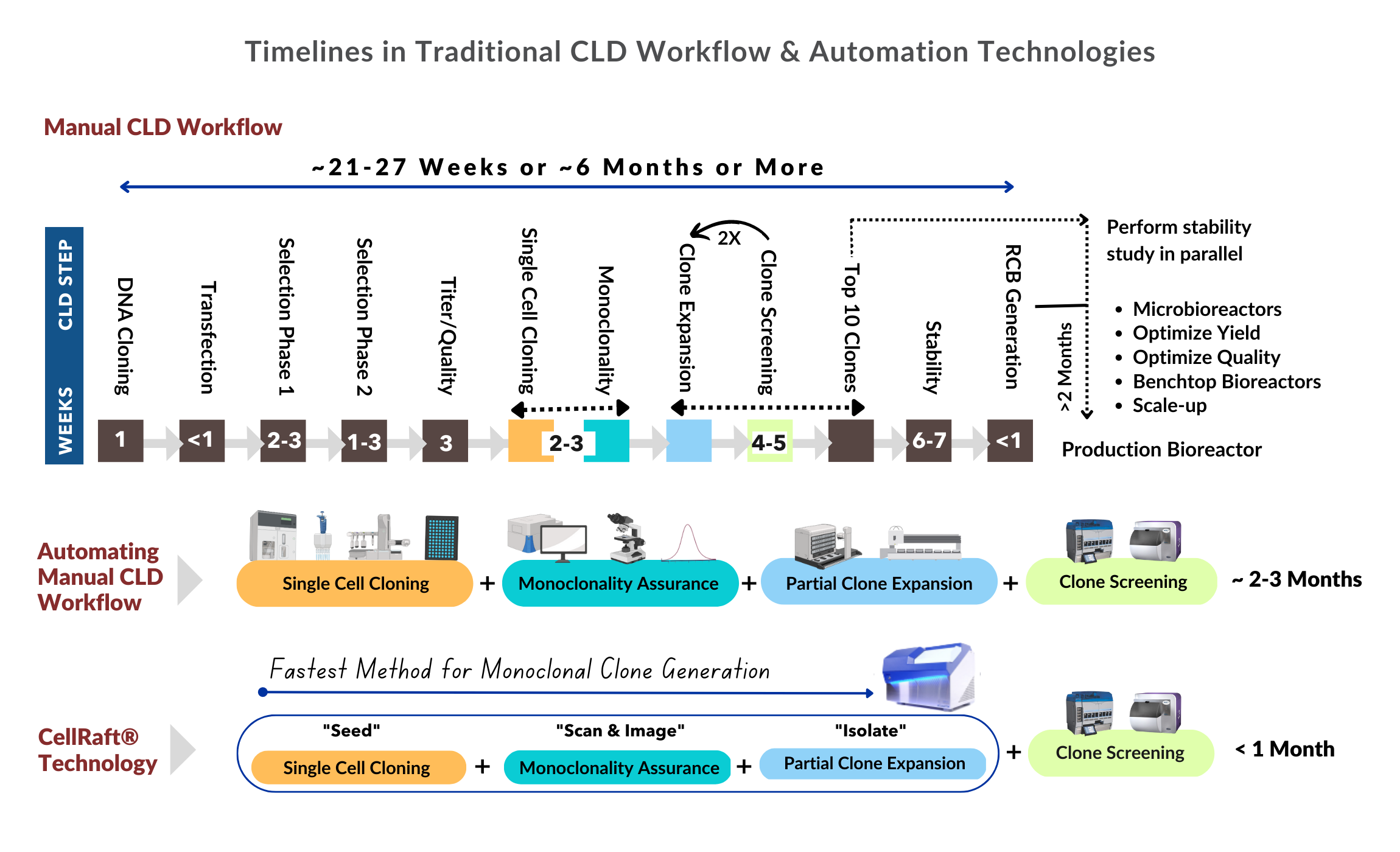
  1. Isolation of a single cell: The first step in single cell cloning is to isolate a single cell from a tissue sample. This can be done using techniques such as flow cytometry or laser capture microdissection. Alternatively, CellRaft® Technology can be used to improve the viability and number of single cell clones generated.
  2. Expansion of the single cell: Once the single cell has been isolated, it is cultured in a medium that allows it to divide and multiply. This process, called expansion, produces a population of cells that are genetically identical to the original single cell.
  3. Characterization of the cell population: The expanded cell population should be characterized to ensure that it is genetically identical and free of contaminants. This may involve DNA fingerprinting, DNA sequencing, or other techniques.
  4. Validation of the cell population: The expanded cell population should be validated to ensure that it is suitable for use in the production of ATMPs. Validation may involve testing the cell population under various conditions to ensure that it produces consistent and reproducible results.
  5. Production of the ATMP: The validated cell population is then used to produce the ATMP, which may involve transfecting the cells with a gene of interest or differentiating the cells into a specific cell type.


Timelines in traditional CLD Workflow Automation Technologies 2300 × 1400 px 12

Learn more about this technology.

Improve viability, clonality, & efficiency – learn more.


REFERENCES
A Risk-based Approach for Cell Line Development, Manufacturing and Characterization of Genetically Engineered, Induced Pluripotent Stem Cell–derived Allogeneic Cell Therapies; Jennifer L. Dashnau, Qiong Xue, Monica Nelson, Eric Law, Lan Cao, Derek Hei. Cytotherapy. Volume 25, Issue 1, January 2023, Pages 1-13. Available online at: https://www.sciencedirect.com/science/article/pii/S1465324922007460.

Transitioning from Preclinical Evidence to Advanced Therapy Medicinal Product: A Spanish Experience; Paloma Gastelurrutia, Cristina Prat-Vidal, Joaquim Vives, Ruth Coll, Antoni Bayes-Genis,Carolina Gálvez-Montón. Frontiers in Cardiovascular Medicine, Volume 8, Article 604434, February 2021. Available online at: www.frontiersin.org.

U.S. Food and Drug Administration: www.fda.gov.



Kap Kumar 2023
Kap Kumar, Ph.D., MBA
Vice President of Strategic Marketing | kkumar@cellmicrosystems.com

Kap Kumar has over 25 years of experience in the life sciences tools and reagents industry. He started in R&D and product development, where he launched products for cell biology and imaging applications. For the last 15 years, Kap has led strategic marketing, market development, and product management for a variety of companies, including Thermo Fisher Scientific (Life Technologies), Danaher (Beckman Coulter Life Sciences), Cell Signaling Technologies, Nexcelom Biosciences, and Avantor-VWR. Kap has diverse experience managing complex portfolios, including instruments, consumables, and reagents, both in early-stage and mature companies. Kap has a Ph.D. in Cell and Molecular Biology from Kent State University, a post-doctorate from Harvard Medical School, and an MBA from Babson College.

Related Posts

Unlocking the Secrets of Antibiotic Resistance: How Shear Flow Systems Are Revolutionizing the Fight Against Superbugs

In the ongoing battle against antibiotic-resistant bacteria, or “superbugs,” scientists are constantly seeking innovative tools to understand the mechanisms of resistance and develop effective treatments. One such groundbreaking technology is shear flow systems, which have emerged as game-changers in microbiological research. By simulating the dynamic conditions of bacteria’s natural environments, such as the human body or food processing, shear flow systems provide unique insights into bacterial behavior and resistance mechanisms that traditional static methods simply cannot achieve. In this blog post, we’ll explore the benefits of using shear flow systems to combat antibiotic resistance and develop next-generation therapies.

Why Shear Flow Systems Are Essential for Studying Antibiotic Resistance

Antibiotic resistance is a complex phenomenon influenced by a variety of factors, including genetic mutations, horizontal gene transfer, and the formation of biofilms. Traditional laboratory methods often fail to capture the dynamic nature of these processes, leading to incomplete or misleading results. Shear flow systems address this limitation by providing a more realistic environment for bacterial growth and interaction. Here’s how:

1. Mimicking Real-World Conditions

In the human body, bacteria are rarely in a static state. They are constantly exposed to fluid flow, such as blood circulation or urine flow, which influences their behavior and resistance mechanisms. Shear flow systems can replicate these conditions, allowing researchers to study how bacteria respond to antibiotics under realistic physiological conditions. This is critical for understanding how resistance develops and persists in vivo. This benefit was recently highlighted in a webinar presented by Dr. Katharina Richter, a microbiology researcher from the University of Adelaide in Australia. Using a high-throughput BioFlux Shear Flow System, Dr. Richter and her team were able to test 3 different methods of superbug treatments under physiological conditions.

2. Studying Biofilm Formation and Resistance

Biofilms—structured communities of bacteria encased in a protective matrix—are a major contributor to antibiotic resistance. Biofilms are notoriously difficult to treat because they shield bacteria from antibiotics and the immune system. Shear flow systems enable researchers to study biofilm formation in real time, observing how bacteria adhere to surfaces, form microcolonies, and develop resistance under flow conditions. For example, using a BioFlux, Dr. Richter was able to leverage high-resolution imaging to obtain unprecedented insight into the biofilm killing and prevention efficacy of a copper and diethyldithiocarbamate (DDC) combination nanoparticles¹. The group is currently investigating the use of a Cu-DDC infused gel that can be injected into wounds that are at high risk for chronic infection, such as hernia.

From Milliliters to Microliters: How a Shear Flow System Can Reduce Sample Requirements for Experiments

Imagine working with a rare patient-derived cell samples. In a traditional setup, you might need 1 milliliter of sample (equivalent to about 20 drops) to run an experiment. With a microfluidic system, you could potentially get the same results with just 10 microliters (about one-half of a single drop). This efficiency opens up entirely new research possibilities, especially in fields where sample availability is a limiting factor.

Whether due to rarity, cost, or difficulty in obtaining samples, the ability to conduct meaningful experiments with limited sample volumes has long been a goal in biological and chemical research. In this quest for more efficient and precise laboratory techniques, microfluidic shear flow systems have emerged as game-changing tools. These systems are not only reducing the amount of sample required for experiments but are also proving their versatility across a wide range of sample types.

Sample volume

Traditional experimental setups, including static and non-microfluidic shear flow, often demand significant amounts of material, which can be both costly and impractical. This is especially true when working with precious or limited samples such as rare biological specimens, expensive chemicals, or newly synthesized compounds. Enter microfluidic shear flow systems – an elegant solution that’s revolutionizing how investigators approach assays. These miniaturized platforms are proving invaluable across multiple disciplines by dramatically reducing sample requirements from milliliters down to microliters, while maintaining experimental integrity. This up to 1000-fold reduction in sample volume is particularly crucial when working with substances, such as patient-derived cells or novel pharmaceuticals. By significantly reducing the volume of reagents and samples needed, these systems cut costs and make experiments more sustainable.

Sample versatility

Equally as impressive as their sample and reagent reduction ability, is the versatility of microfluidic shear flow systems to adapt to different types of samples.

  • Human Samples: Researchers can study cellular behavior under controlled shear stress, mimicking physiological conditions such as blood or saliva flow.
  • Nanomaterials: The precise fluid dynamics in these systems allow for the uniform dispersion and manipulation of nanoparticles, enabling high-resolution studies in materials science.
  • Microbes: Continuous fluid flow more closely represents the natural environments of many microbes, such as plumbing, food processing, and chronic wounds.

The ability to work with multiple sample types in the same system allows researchers to explore different experimental conditions without the need for multiple specialized setups. By precisely controlling channel geometries and flow rates, researchers can subject different sample types to well-defined shear conditions, enabling detailed studies of cellular mechanics, protein interactions, and material properties.

Microfluidic…
Single Cells: Lab Tune Sung to the Rhythm of "Jingle Bells"

By Jessica Hartman, Ph.D.

Toiling in the lab

With a deadline on its way

To the scope I go

On a Saturday

I don’t see a clone

I’ve looked for one that’s right

What misery these data bring,

I’ll have to look all night,

Oh! Single cells, single cells, dilutions aren’t the way

Oh, I wish I had an AIR to pick a clone, to-da-ay!

Single cells, single cells, I need a better way

Use a Raft to grow a clone or be sad this holiday!

Use a Raft to grow a clone or be sad this holiday!

Now it’s getting late

My spirits are so low

My PI will hate

If my cells don’t grow

My eyesight’s getting dim

A clone I cannot see

My chances are so slim

I need CellRaft Cytometry

Oh! Single cells, single cells, sorters aren’t the way

Oh, I wish I had an AIR to pick a clone, to-da-ay!

Single cells, single cells, I need a better way

Use a Raft to grow a clone or be sad this holiday!

Use a Raft to grow a clone or be sad this holiday!

Oh! Single cells, single cells, dispensers aren’t the way

Oh, I wish I had an AIR to pick a clone, to-da-ay!

Single cells, single cells, I need a better way

Use a Raft to grow a clone or be sad this holiday!

Use a Raft to grow a clone or be sad this holiday!


 “Single Cell” vocalist: Virginia Laurie

Jessica Hartman, Ph.D.Senior Director of Product Applications | jessica.hartman@cellmicrosystems.comDr. Jessica Hartman has a B.S in Biology from the University…