

You never forget the first time you thaw a vial of induced pluripotent stem cells. Standing in the lab, holding a precious vial of cells, reading, and re-reading the protocols, and crossing your fingers that you don’t kill the cells, you gingerly hold that vial in a waterbath like it is a rare diamond. In almost 20 years of cell culture experience, no cell type has inspired as much fear and awe in my scientific heart than that vial of iPSCs.
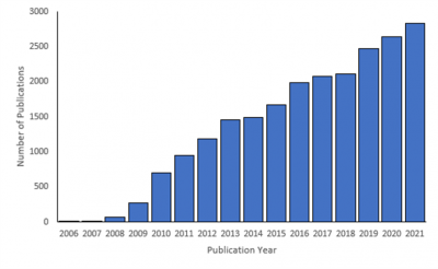
Figure 1. The number of publications referencing the term “iPSC” in PubMed since 2006.
Since their discovery in 2006 by Takahashi and Yamanaka, induced pluripotent stem cells (iPSCs) have seen a meteoric rise in their use throughout academic and industrial research, and to date, there are over 21,000 publications in PubMed referencing them (Figure 1). Generated by reprogramming terminally differentiated somatic cells with four key transcription factors (Oct4, Sox2, KLF4, and c-myc), human iPSCs have the potential to unlimitedly proliferate and differentiate into all of the key cell types in the human body. These cells hold promise for a myriad of therapeutic research areas, including gene therapy, regenerative medicine, and personalized medicine. In addition, they alleviate many of the ethical concerns and sourcing issues associated with pluripotent cells such as human embryonic stem cells. And yet, despite countless research, rapidly advancing protocols, and increased access for researchers at all levels, iPSCs remain a challenging enigma that pose some key limitations for their utility in therapeutic research.
So, what is it about iPSCs that can strike fear into the heart of an experienced bench scientist?
To overcome these bottlenecks and challenges to iPSC growth, there are a variety of specialized medias, additives, and tissue culture dish coatings that are meant to improve cell survival in vitro. However, despite these options, cell viability is often compromised, and the phenotype of the line can become unstable with prolonged culture. In addition, iPSC lines require constant maintenance, necessitating daily media changes and manual manipulation to remove areas of differentiation or to isolate desired clones for further study.
The improvement of iPSC culture conditions and the development of automated iPSC workflows have the potential to increase the utility, ease, and throughput of these workflows, thereby accelerating the use of iPSCs in personalized medicine and drug discovery.
A novel solution that enables this possibility is the CellRaft® Technology from Cell Microsystems. Briefly, the key features of the technology that are beneficial for iPSC workflows include:
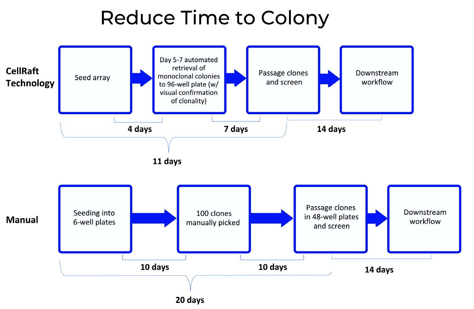
Figure 2: Compared to a manual limiting dilutionA statistical cloning approach where cells are diluted to low density to “hopefully” seed one cell per well; commonly used but can be low-efficiency for difficult-to-clone lines. More process, clones were able to be passaged and screened nine days earlier using CellRaftA microscale polystyrene growth surface within an array used to spatially segregate cells/colonies while maintaining shared media access, enabling imaging over time and targeted isolation. More Technology. In addition, less hands-on time was required with CellRaftA microscale polystyrene growth surface within an array used to spatially segregate cells/colonies while maintaining shared media access, enabling imaging over time and targeted isolation. More Technology as the system is able to automatically retrieve colonies and move them to 96-well plates.
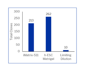
To determine whether these key features could alleviate bottlenecks hindering iPSC workflows, we compared clonal iPSC development using CellRaftA microscale polystyrene growth surface within an array used to spatially segregate cells/colonies while maintaining shared media access, enabling imaging over time and targeted isolation. More Technology to traditional limiting dilutionA statistical cloning approach where cells are diluted to low density to “hopefully” seed one cell per well; commonly used but can be low-efficiency for difficult-to-clone lines. More. Using this method, we were able to generate over 200 single cell derived iPSC clones on a single consumable, compared to 10 clones in a single 96 well plate (Figure 2). This workflow required 1000X less iPSC coating and 2000X less media per cell screened. In addition, the image acquisition over time allows for the detailed assessment of monoclonalityEvidence that a colony/line originated from one cell; often supported by time-course imaging and traceability. More, ensuring that successful clones are not heterogeneous and eliminating the need for downstream clonal characterization and shortening the time to use in downstream applications (Figure 3).
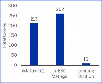
Figure 3: CellRaftA microscale polystyrene growth surface within an array used to spatially segregate cells/colonies while maintaining shared media access, enabling imaging over time and targeted isolation. More AIR vs limiting dilutionA statistical cloning approach where cells are diluted to low density to “hopefully” seed one cell per well; commonly used but can be low-efficiency for difficult-to-clone lines. More for monoclonal iPSC development. iPSCs were seeded on either a CellRaft ArrayConsumables containing thousands of microwells (containing CellRafts) for spatial segregation, imaging, and isolation of single cells, monoclonal colonies, or 3D cultures. More coated with iMatrix-511 (Matrixome) or h-ESC Matrigel (Corning) or on iMatrix-511 coated 96 well plates for limiting dilutionA statistical cloning approach where cells are diluted to low density to “hopefully” seed one cell per well; commonly used but can be low-efficiency for difficult-to-clone lines. More. Colony formation was monitored using the CellRaftA microscale polystyrene growth surface within an array used to spatially segregate cells/colonies while maintaining shared media access, enabling imaging over time and targeted isolation. More AIR or manual observation.
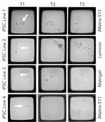
Figure 4: Track and trace of iPSC clone formation on the CellRaftA microscale polystyrene growth surface within an array used to spatially segregate cells/colonies while maintaining shared media access, enabling imaging over time and targeted isolation. More AIR. Four different iPSC cell lines were seeded on CellRaftA microscale polystyrene growth surface within an array used to spatially segregate cells/colonies while maintaining shared media access, enabling imaging over time and targeted isolation. More arrays on one of three coatings (iMatrix-511, h-ESC Matrigel, or Laminin). The arrays were serially scanned starting 4 hours post-cell seeding and every 24 hours after to monitor clone formation.
Ultimately, the ability to screen tens of thousands of iPSCs on a single consumable using a fully integrated software and instrument platform to identify and isolate iPSC clones of interest has the potential to greatly increase the utility of iPSCs, improve iPSC characterization, and decrease the time to cell generation and therapeutic discovery.
For more on iPSC lines, view Dr. Hartman’s RaftNote: Taking the fear out of iPS cell line development.


Dr. Jessica Hartman has a B.S in Biology from the University of Virginia, a Ph.D. from Duke University in Molecular Cancer Biology and postdoctoral training in Biochemistry and Cancer Biology at Baylor College of Medicine and Duke University, respectively. She has previously served in Director-level roles, managing bioscience research and development for biotechnology companies. At Cell Microsystems, Dr. Hartman’s role is to lead the development of new and streamlined workflows using the CellRaftA microscale polystyrene growth surface within an array used to spatially segregate cells/colonies while maintaining shared media access, enabling imaging over time and targeted isolation. More Technology and its associated products.




By Jessica Hartman, Ph.D.
Toiling in the lab
With a deadline on its way
To the scope I go
On a Saturday
I don’t see a clone
I’ve looked for one that’s right
What misery these data bring,
I’ll have to look all night,

Oh! Single cells, single cells, dilutions aren’t the way
Oh, I wish I had an AIR to pick a clone, to-da-ay!
Single cells, single cells, I need a better way
Use a Raft to grow a clone or be sad this holiday!
Use a Raft to grow a clone or be sad this holiday!
Now it’s getting late
My spirits are so low
My PI will hate
If my cells don’t grow
My eyesight’s getting dim
A clone I cannot see
My chances are so slim
I need CellRaftA microscale polystyrene growth surface within an array used to spatially segregate cells/colonies while maintaining shared media access, enabling imaging over time and targeted isolation. More Cytometry
Oh! Single cells, single cells, sorters aren’t the way
Oh, I wish I had an AIR to pick a clone, to-da-ay!
Single cells, single cells, I need a better way
Use a Raft to grow a clone or be sad this holiday!
Use a Raft to grow a clone or be sad this holiday!
Oh! Single cells, single cells, dispensers aren’t the way
Oh, I wish I had an AIR to pick a clone, to-da-ay!
Single cells, single cells, I need a better way
Use a Raft to grow a clone or be sad this holiday!
Use a Raft to grow a clone or be sad this holiday!


“Single Cell” vocalist: Virginia Laurie