In the 1990s, if you wanted to study the activity of ionotropic receptors across the membrane of cells, your best bet was learning how to patch clamp. This technique requires careful positioning of a glass pipette filled with a saline solution that mimics the internal ionic concentration in the cytoplasm close to a single cell. Without destroying the cell, careful suction creates a giga-Ohm (GΩ) seal and, later, disruption of the membrane patch allows electrical access. This manual patch clamp (MPC) technique, the so-called “Gold Standard” is time-consuming, tedious, and requires months of training to achieve any results and even longer for reliable results.
Although it is still held as a benchmark by electrophysiologists, with the focus of today’s world being on rapid deliverables in drug screening, manual patch clamp simply doesn’t cut it. Of course, in some conditions, manual patch clamp is still the only way to study ionic channel activities, but the road to automation is getting wider and easier to navigate day by day.
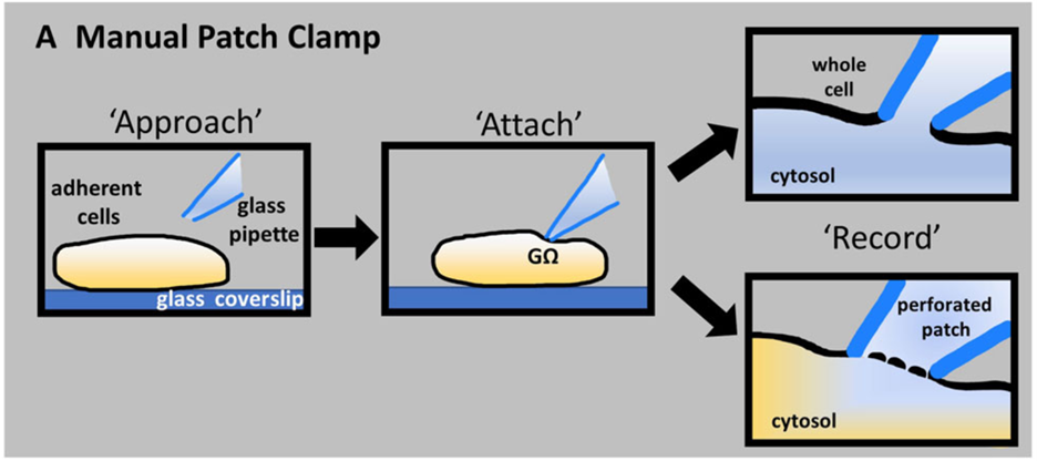
Figure 1: Representation of the manual patch clamp technique where a glass pipette is brought close to touch the cell to form a giga-Ohm (GΩ) seal. Subsequently, gentle suction ruptures the patch of membrane to form a whole cell patch or added compounds allow for a perforated patch. D Bell & M Dallas, 2017
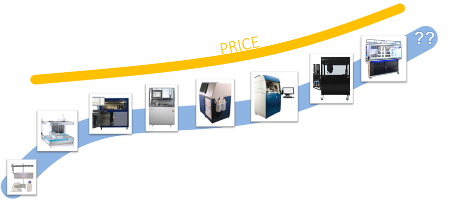
In the late 1990s, Automated Patch Clamp systems (APCs) began to appear and quickly supplemented or even replaced MPC in industrial situations. With the popularity of recombinant homogenous cell lines that overexpressed target proteins, drug screening against ion channels quickly shifted from MPC (~20-30 data points per day) to automated systems (~100-1000s data points per day). In addition, APCs greatly reduced the technical requirements for initiating these studies and provided a high-quality alternative to MPC. APCs not only sped up drug screening and safety pharmacology but also enabled access to the patch clamp technology in environments that would have otherwise utilized less definitive alternatives. As the need for greater throughput increased, the relative complexity, and hence the operational and ownership costs of APCs, increased. Although APCs were effective, the exorbitant cost limited their appeal in academic and small lab settings, making MPC systems the de facto alternative in these environments.
Figure 2: Representation of APC (from a single channel to 384/768) over the years with increasing recording sites and increasing prices.
It is now established that APCs, with their large number of recording sites, can increase ion channel research throughput, but how can the cost of ownership go down to a point of academic and small lab accessibility? The common feature among most APCs that makes them cost-prohibitive is their dependence on complicated and expensive liquid handlers to provide cell and solution delivery. However, in 2011, IonFlux, an alternative microfluidic-based APC system, was released. Utilizing lateral patch clamp, with in-plate liquid delivery by microfluidic channels, IonFlux APCs leverage pneumatic pumps to precisely drive and control solution exchange.
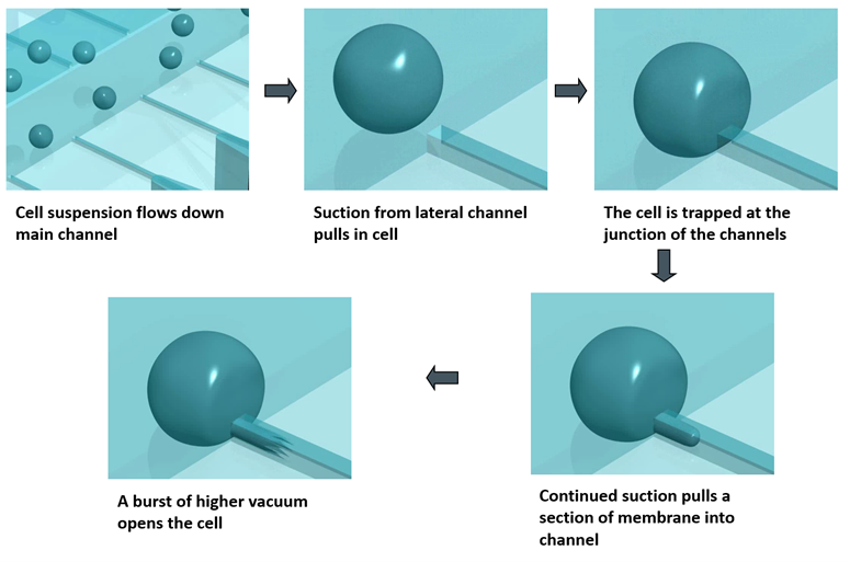
Figure 3: Lateral patch clamp provides the means to leverage more cost-effective microfluidics.
The specialized IonFlux plates are SBS standard-sized plates with intricate microfluidic channels embedded into the bottom. The channels are divided into experimental patterns with dedicated wells for cell delivery, cell trapping/patching, and individual compound delivery. Each 12-well pattern is isolated and devoted to its own experiment, eliminating cross-contamination and drastically reducing sample waste.
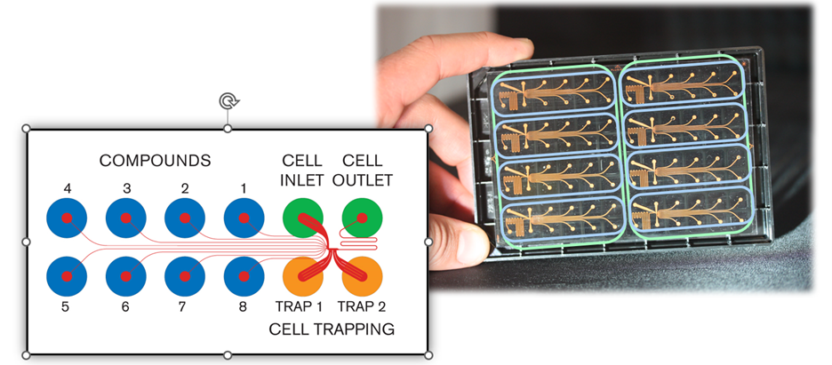
Figure 5: IonFlux plates (IonFlux 16 plate to the right) are divided into experimental patterns. Each experimental pattern has a dedicated cell inlet, cell outlet two trapping wells that contain intracellular solutions, and 8 dedicated compound wells (example to the left). Each compound travels to the experiment area using its own dedicated microfluidic conduit making high throughput parallel assays possible.
This microfluidic technology drastically reduces the cost of owning and operating an APC system, but can it effectively replace manual patch clamp? In certain conditions, yes.
Four major factors that affect the replacement of MPC with APC:
“Finally, our studies have demonstrated that microfluidic patch clamp methods are a suitable substitute to conventional methods for screening GABAA receptor modulators. We found that the microfluidic approach had several advantages over conventional methods. First, the apparatus is straightforward to use, and unlike conventional methods, it does not require prolonged training. Second, the miniaturized set-up means that drug or biological fluid use is reduced by 2–10-fold per data point. This is especially useful if screening small volumes of human samples, expensive therapeutics, or screening difficult to synthesize compounds. In fact, while each determination may need only 50 µL of drug to set up the assay, after the experiment, 90% may be reclaimed for subsequent analysis. “ Kaplan et al, 2023
Automated patch clamp technology represents a significant advancement in electrophysiological research. APCs have greatly improved since their advent and when combined with the use of recombinant cell lines, many APC systems provide exponentially greater throughput, reliable high-quality recordings, and similar or better flexibility compared to manual patch clamp. The cost of automated patch clamp systems varies based on factors such as the number of cells recorded simultaneously, features, and additional accessories. Although a percentage of these costs are offset by increased efficiency and data quality, IonFlux APCs leverage microfluidics to drive the cost down to a point of affordability that is feasible for both industrial and academic settings. Together, these factors have made automated patch clamp systems viable alternatives to manual patch clamp systems in drug screening environments.

Ali Yehia has a Ph.D. in Cardiac Electrophysiology from McGill University. He has been involved in ion channel screening using Automate Patch Clamp since 2006. He joined Fluxion Biosciences in 2012, where he developed ion channel assays and IonFlux Mercury systems. He was the Chief Scientific Officer at Fluxion before moving to Cell Microsystems as the new Senior Director of the IonFlux product.


In the ongoing battle against antibiotic-resistant bacteria, or “superbugs,” scientists are constantly seeking innovative tools to understand the mechanisms of resistance and develop effective treatments. One such groundbreaking technology is shear flow systems, which have emerged as game-changers in microbiological research. By simulating the dynamic conditions of bacteria’s natural environments, such as the human body or food processing, shear flow systems provide unique insights into bacterial behavior and resistance mechanisms that traditional static methods simply cannot achieve. In this blog post, we’ll explore the benefits of using shear flow systems to combat antibiotic resistance and develop next-generation therapies.
Antibiotic resistance is a complex phenomenon influenced by a variety of factors, including genetic mutations, horizontal gene transfer, and the formation of biofilms. Traditional laboratory methods often fail to capture the dynamic nature of these processes, leading to incomplete or misleading results. Shear flow systems address this limitation by providing a more realistic environment for bacterial growth and interaction. Here’s how:
1. Mimicking Real-World Conditions
In the human body, bacteria are rarely in a static state. They are constantly exposed to fluid flow, such as blood circulation or urine flow, which influences their behavior and resistance mechanisms. Shear flow systems can replicate these conditions, allowing researchers to study how bacteria respond to antibiotics under realistic physiological conditions. This is critical for understanding how resistance develops and persists in vivo. This benefit was recently highlighted in a webinar presented by Dr. Katharina Richter, a microbiology researcher from the University of Adelaide in Australia. Using a high-throughput BioFlux Shear Flow System, Dr. Richter and her team were able to test 3 different methods of superbug treatments under physiological conditions.
2. Studying Biofilm Formation and Resistance
Biofilms—structured communities of bacteria encased in a protective matrix—are a major contributor to antibiotic resistance. Biofilms are notoriously difficult to treat because they shield bacteria from antibiotics and the immune system. Shear flow systems enable researchers to study biofilm formation in real time, observing how bacteria adhere to surfaces, form microcolonies, and develop resistance under flow conditions. For example, using a BioFlux, Dr. Richter was able to leverage high-resolution imaging to obtain unprecedented insight into the biofilm killing and prevention efficacy of a copper and diethyldithiocarbamate (DDC) combination nanoparticles¹. The group is currently investigating the use of a Cu-DDC infused gel that can be injected into wounds that are at high risk for chronic infection, such as hernia.
The study of biofilm communities presents unique challenges in experimental design and analysis. While mixed bacterial populations offer fascinating insights into microbial interactions and community dynamics, accurately characterizing and investigating these complex systems has previously required sophisticated and tedious technical approaches. Here, we will discuss advances in technology that have helped to simplify and streamline polymicrobial investigations.
Throughout the last decade, a trend for growing and assaying biofilms under shear flow has gained traction among microbiologists. The rationale for investigating biofilms under flow is multi-dimensional. A recent Cell Microsystems survey asked why microbiologists are using shear flow found that the top responses were:
Of the scientists surveyed, 40% indicated that they use a BioFlux Shear Flow System. This system provides unique advantages over other methods to induce shear stress, such as slide-based parallel plate flow chambers and benchtop rockers.
BioFlux uses microfluidic channels embedded onto the bottom of standard-sized 6, 24, or 48-well plates to create isolated and independent flow chambers where user-defined wall shear stress is initiated and maintained by digitally controlled air pressure. This not only simplifies the setup and experimental processes but also ensures that experiments are contained and do not contaminate the shear flow system.
Although shear flow has been thoroughly demonstrated to improve the biological relevance of biofilm investigations1, an often overlooked aspect of biofilm physiology is the significant role that multicellular aggregates play in biofilm formation2. When investigating Pseudomonas aeruginosa, it has been demonstrated that multicellular aggregates lead to faster biofilm formation and enhanced resistance to antimicrobials and immune responses3. In addition, multispecies microbial aggregates have been shown to dominate single cells in human saliva4. Together, these data show that aggregates are important areas of investigation because they are more likely to represent physiological biofilm formation and growth.