Is there life after graduate school if you decide not to become an academic faculty member?
 Despite the taboo nature of this question, it is one most scientists face when they decide to pursue a career in science.When contemplating what kind of career I wanted after my postdoc, I knew I wanted to remain in the science field but not necessarily be at the bench full-time. I loved troubleshooting experiments and mentoring others but grew tired of repeating the same experiments day in and day out. I also found that the traditional academic mindset of only focusing on one project wasn’t for me – there’s an infinite amount of fascinating science being done, and I’m interested in all of it!
Therefore, a significant portion of my postdoc was spent working with my institution’s career development office to explore options that aligned with my goals. I learned that there are many non-traditional career opportunities for Ph.D. Scientists. Some of these pathways I had heard about before, such as being an industry bench scientist, teaching in a university setting, or working for a government agency. I was also introduced to roles I hadn’t contemplated before, including Regulatory Affairs, Patent Law, and Field Applications. While reading the job postings for these types of positions offered insight, I discovered that the best way to truly understand these roles was to conduct informational interviews with scientists who had left academia for these careers. After many LinkedIn messages and Zoom calls, one role ticked all the boxes: a Field Application Scientist (FAS).
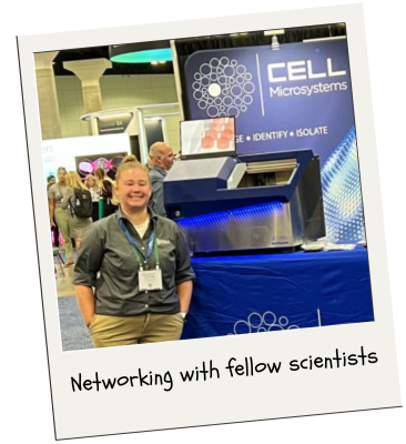 After nearly a year as an FAS, which I like to describe as a traveling troubleshooting scientist, I can say that I have found the perfect fit. As an FAS at a fast-paced instrumentation biotech company, my days and weeks are never the same, and I get to wear many hats. Some weeks, I have the flexibility to work from home and connect with customers, addressing their inquiries about experiments and providing guidance on effectively using our tools. This also gives me time to familiarize myself with the latest scientific literature, enabling me to identify new challenges for our technology to solve.
However, my favorite part of this job is being on the road and engaging with a diverse group of scientists. Whether it is participating at scientific conferences, manning our booth at tradeshows, or conducting demonstrations at potential customer sites and installations with new customers, I love being out in the field and sharing our technology with others.
That technology, which includes the CellRaft® Arrays and CellRaftA microscale polystyrene growth surface within an array used to spatially segregate cells/colonies while maintaining shared media access, enabling imaging over time and targeted isolation. More AIR® System, allows for imaging, identification, and isolation of single cells, monoclonal colonies, or organoids of interest. The CellRaft ArrayConsumables containing thousands of microwells (containing CellRafts) for spatial segregation, imaging, and isolation of single cells, monoclonal colonies, or 3D cultures. More provides spatial segregation of cells while allowing them to grow in contiguous media (Check out my previous blog post to see why that’s important!). Because most cells don’t like to be alone in a well, we seed the cells and let them grow into a small colony. This means that our demos are a week long!
After a year of traveling coast-to-coast and conducting numerous demonstrations, I have gathered some valuable insights:
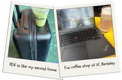
1. Binging on caffeine
Whether it’s a quick coffee or matcha at the airport or finding a fun coffee shop to analyze data in – caffeine is a must, especially when traveling back and forth between time zones!
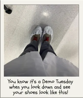
2. Gowning up as an FAS
I do occasionally miss being at the bench, so I love it when I get to gown and glove up and demonstrate our instrument at customer sites! The best part of my job, in my opinion, is going into a lab and showing scientists how our instrument can help with their research. Many scientists who request to demo our instrument struggle to obtain monoclonal colonies from difficult edited cells. With our system, they are able to see their cells at the single cell stage and watch them grow into colonies before they isolate – this is unique in that they have the ability to choose the clones they want and when they want them. It’s so rewarding to hear, “Wow, I’ve never seen my cells like this before!” when observing them go from one cell to two cells to a small colony.
3. Taking time to explore
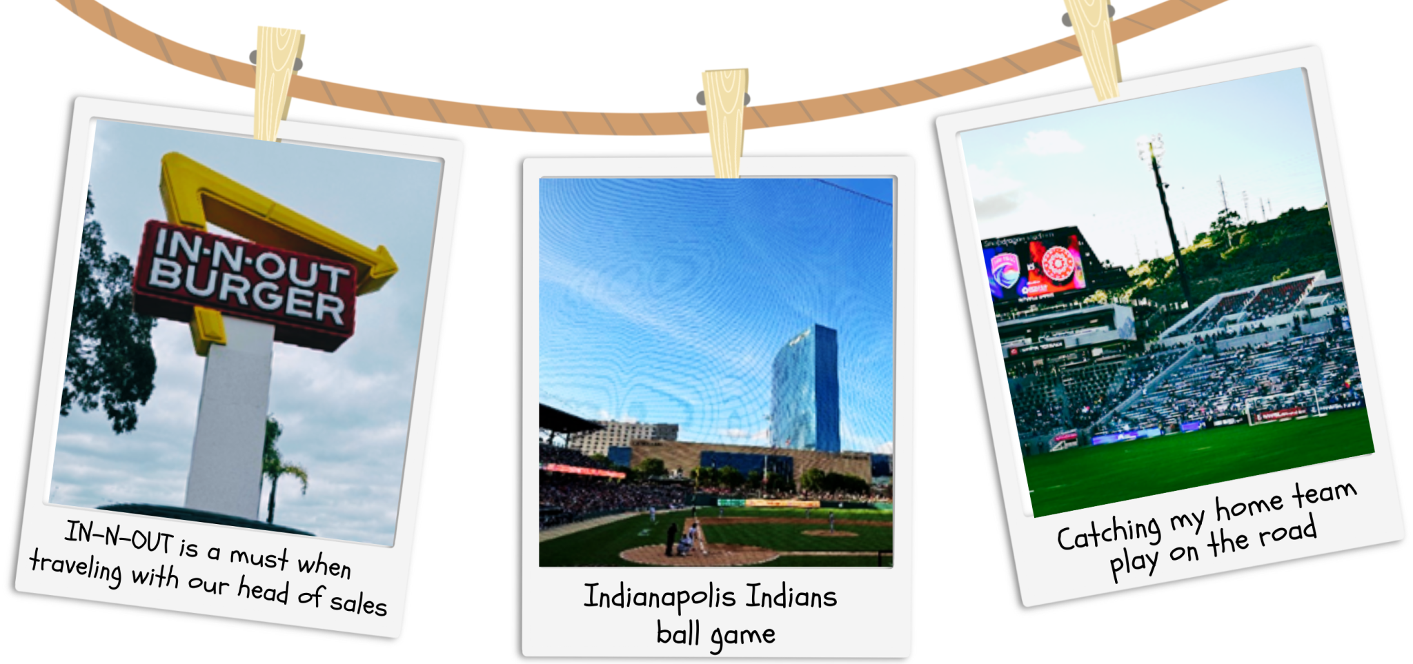
Being on the road so often and juggling demos with current customer needs can be challenging. However, one thing I’ve learned from my sales team is the importance of taking time to explore whatever new city I’m in and doing at least one thing I enjoy. For example, one colleague makes it a point to get IN-N-OUT burgers whenever we’re in California, while another enjoys finding minor-league baseball games to attend. As for me, my favorite ways to explore are seeking out fun coffee shops (as mentioned above) and attending pro soccer matches.
 4. Keeping in contact with my team
Being in the field so often and working remotely across the country from the home office can sometimes feel isolating. I find it’s super important to communicate and keep up with my coworkers. Staying in touch is definitely a must, whether it’s through weekly Teams calls with my boss and counterpart on the East Coast, the convenience of messaging one of our awesome researchers and software experts whenever I have a customer question, or virtually joining in on our company’s gamers chat.
Trying to find a job after grad school/postdoc can be daunting, especially when academia is often presented as the default route. However, alternative scientific careers can be equally rewarding! As an FAS, I get to travel the world helping scientists further their research and teaching about the capabilities of CellRaftA microscale polystyrene growth surface within an array used to spatially segregate cells/colonies while maintaining shared media access, enabling imaging over time and targeted isolation. More Technology. The best advice I could give to those looking to jump the academic ship is to reach out to those who have already done it and learn about the career paths available. That being said, if you’re interested in hearing more about being an FAS or about the CellRaft AIR SystemAn all-in-one platform for imaging, identifying, and isolating viable single cells, colonies, or organoids using CellRaft Arrays and software-guided selection with CellRaft Cytometry. More, feel free to reach out via LinkedIn!
Dr. Schoepke received post-doctoral training in translational breast cancer research at Baylor College of Medicine, obtained a Ph.D. in Pharmacology and Physiology from Saint Louis University, and holds a Bachelor of Science in Molecular and Cellular Biology with a minor in Chemistry from the University of Illinois at Urbana-Champaign. Her background is in Nuclear Receptor pharmacology, testing novel drugs in 2D and 3D cell-based assays of cancer and in vitro models of metabolic disease and exercise. Dr. Schoepke is currently a Field Application Scientist at Cell Microsystems who focuses on demonstrating and training new customers on the CellRaft® AIR System as well as troubleshooting novel single cell workflows.