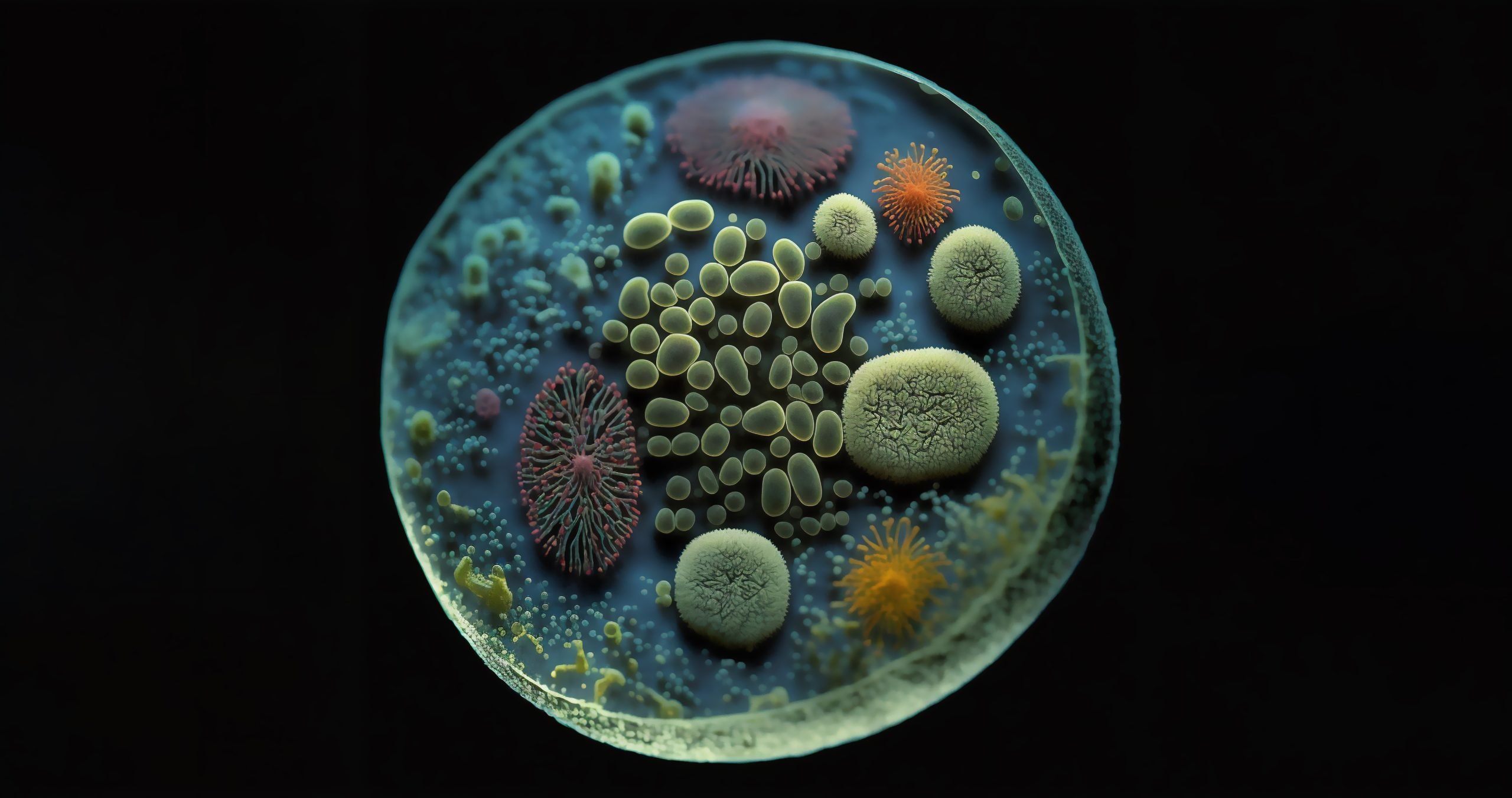
Physiological relevance versus reproducibility — As scientists, we all recognize the limitations of developing assays using 2D immortalized cell lines and thus have dedicated our careers to developing cellular models that more closely recapitulate in vivo biology. The reality is, with each step we take down the path of complexity—primary cells, co-culture, 3D models—we also introduce new challenges associated with heterogeneity and variability that must be addressed to generate robust and reproducible data.
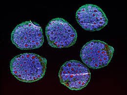
Scientists in the fields of research and therapeutic discovery have recognized the potential of organoid models to transform and accelerate basic and translational research programs, regenerative medicine, and preclinical pipelines. These stem-cell derived 3D models offer greater physiological relevance over other models because:
Despite these promising advantages of organoids, there are a host of technical challenges associated with culture methods, as well as analytical limitations, that prevent organoids from being more widely adopted as high-throughput cellular models early in the drug discovery pipeline.
Common pain points in organoid culture
There are two main reasons organoid cultures present challenges in cell-based assays 1) establishment of reproducible cultures, and 2) population heterogeneity. Most organoid models rely on extracellular matrix (ECM) to form 3D structures. Seeding organoids into ECM droplets, or domes, is an incredibly laborious process that relies on precise pipetting, temperature control, and expert users. Even with a developed SOP and well-trained hands, these experimental nuances cause a spectrum of variability between wells, such as organoid number, dome size and placement in the well. In addition to well-to-well variability, organoid domes containing 10s to 100s of organoids are heterogenous themselves, which presents challenges in imaging and analysis, such as (Figure 1):
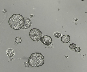
Figure 1. Organoids grown in a dome of ECM have large population variability, including size and viability inconsistencies, require multifocal imaging, and are limited to pooled readouts.
Ultimately, data generated from these heterogenous populations of organoids often mask or misrepresent the biological response. Because of these challenges, establishing reproducible organoids for both inter- and intra-experiment consistency is perhaps the biggest barrier preventing these models from being useful for moderate-to-high throughput screening assays.
One approach to evaluating population heterogeneity is to use imaging and advanced analysis to evaluate individual organoid responses. To interrogate single organoid responses to a drug or other perturbation, a researcher would need hi-tech imaging instrumentation and analytical tools to parse out single-organoid imaging data from a population of organoids. Unfortunately, if further evaluation of individual organoids in the population is necessary, your experiment has reached the end of the line—the organoids you’ve spent a great deal of time and effort to evaluate using image analysis are stuck, along with all the other organoids you may or may not care about, inside the ECM dome.
What if there was a way to escape the dome?
Outside the Dome: A novel culture method for organoids
Improving the scalability, efficiency, and reproducibility of organoid models for screening applications is one of the biggest challenges researchers face in using these complex 3D models for their research and discovery programs. The CellRaft® Technology offers a unique solution to many of the pain points researchers face in their organoid workflows. Using the core technology, the CellRaft® Array, and the CellRaftA microscale polystyrene growth surface within an array used to spatially segregate cells/colonies while maintaining shared media access, enabling imaging over time and targeted isolation. More AIR® System, users can:
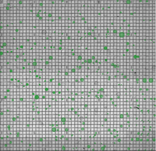
Figure 2. Organoids cultured on the CellRaft ArrayConsumables containing thousands of microwells (containing CellRafts) for spatial segregation, imaging, and isolation of single cells, monoclonal colonies, or 3D cultures. More. Field-of-view images, acquired on the CellRaft AIR SystemAn all-in-one platform for imaging, identifying, and isolating viable single cells, colonies, or organoids using CellRaft Arrays and software-guided selection with CellRaft Cytometry. More, have been stitched together to generate a representative image of the entire CellRaft ArrayConsumables containing thousands of microwells (containing CellRafts) for spatial segregation, imaging, and isolation of single cells, monoclonal colonies, or 3D cultures. More. Green circles highlight every CellRaftA microscale polystyrene growth surface within an array used to spatially segregate cells/colonies while maintaining shared media access, enabling imaging over time and targeted isolation. More in the array containing a single organoid that can be imaged, analyzed, and isolated for downstream use.
Use Case Data
To demonstrate the ability of the CellRaft AIR SystemAn all-in-one platform for imaging, identifying, and isolating viable single cells, colonies, or organoids using CellRaft Arrays and software-guided selection with CellRaft Cytometry. More to produce intra-assay consistency for organoid screening assays, we grew, serially imaged, and used software tools to identify organoids of interest for isolation. To decrease the size heterogeneity of the downstream screening assay, software tools were used to identify two populations of organoids. The first population was created to identify CellRafts with single organoids, irrespective of size, and the second was created to identify single organoids ranging from 300-500 microns in diameter (Figure 3). CellRafts with organoids of interest were isolated into 96-well plates to generate variable and size-selected plates for downstream drug toxicity screening assays (Figure 4). For detailed experimental design and resulting drug toxicity data see our Organoid RaftNote.
In summary, the data demonstrate that using the CellRaft AIR systemAn all-in-one platform for imaging, identifying, and isolating viable single cells, colonies, or organoids using CellRaft Arrays and software-guided selection with CellRaft Cytometry. More we were able to reduce variability in replicate wells using size-selected organoids resulting in dose-response curves that could be translated to ED50 data, which was not possible from the parallel assay with variable-sized organoids. These data highlight the importance of well-to-well consistency, and the ability of CellRaftA microscale polystyrene growth surface within an array used to spatially segregate cells/colonies while maintaining shared media access, enabling imaging over time and targeted isolation. More Technology to provide a solution to this challenge of organoid screening assays.
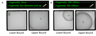
Figure 3. Select organoids for isolation based on phenotypic and morphologic characteristics to generate intra- and inter- assay consistency. To demonstrate the importance of organoid size in single-organoid screening assays, populations were created to represent variable-sized organoids greater than 50um (A), and the second was selected for organoids ranging from 300-500um in diameter.
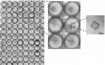
Figure 4. The CellRaft AIR SystemAn all-in-one platform for imaging, identifying, and isolating viable single cells, colonies, or organoids using CellRaft Arrays and software-guided selection with CellRaft Cytometry. More performs automated isolation of CellRafts containing pre-selected organoids of interest into 96-well plates for downstream use, such as drug screening.
Lessons Learned
Using the CellRaft AIR SystemAn all-in-one platform for imaging, identifying, and isolating viable single cells, colonies, or organoids using CellRaft Arrays and software-guided selection with CellRaft Cytometry. More, we have developed workflows and generated data that demonstrate widespread organoid applications. Here, we’ve introduced the key features, such as reliable temporal imaging of hundreds of individual organoids, software-guided selection of organoids of interest, and automated isolation of single organoids for downstream use, that enable efficient and reproducible organoid screening assays. To learn more about how the CellRaftA microscale polystyrene growth surface within an array used to spatially segregate cells/colonies while maintaining shared media access, enabling imaging over time and targeted isolation. More technology can transform organoid workflows, visit our webpage and follow our blog for future organoid content.
Learn more about CellRaftA microscale polystyrene growth surface within an array used to spatially segregate cells/colonies while maintaining shared media access, enabling imaging over time and targeted isolation. More Technology.

Dr. Stern obtained a Ph.D. and Master of Science in Physiology from North Carolina State University and a Bachelor of Science in Animal Science. Her background is in comparative physiology, with a concentration in cell biology and developing 2D and 3D in vitro models for drug and toxicological studies. Dr. Stern is currently a scientist on the Product Applications team at Cell Microsystems where she focuses on developing novel 2D and 3D cellular workflows using the CellRaftA microscale polystyrene growth surface within an array used to spatially segregate cells/colonies while maintaining shared media access, enabling imaging over time and targeted isolation. More Technology and provides customer training and support for 3D workflows using the CellRaft AIR SystemAn all-in-one platform for imaging, identifying, and isolating viable single cells, colonies, or organoids using CellRaft Arrays and software-guided selection with CellRaft Cytometry. More.