

Monoclonal cell lines are a powerful tool for biomedical research and drug development. The development and use of monoclonal cell lines have been accelerated due to the advent of breakthrough gene editing technologies such as CRISPR-Cas9. Due to their ease of use and amenability to single cell cloning, there are several well-established and recognizable cell lines that are commonly used to create monoclonal colonies, including the CHO and HEK293 lines. However, primary cells such as induced pluripotent stem cells (iPSCs), or immortalized cancer cell lines have proven more challenging. Historically, monoclonal cell lines have been developed using traditional methods, such as flow cytometry or manual limiting dilutionA statistical cloning approach where cells are diluted to low density to “hopefully” seed one cell per well; commonly used but can be low-efficiency for difficult-to-clone lines. More. Although widely utilized, these methods present both technical and biological challenges, and technologies such as the CellRaftA microscale polystyrene growth surface within an array used to spatially segregate cells/colonies while maintaining shared media access, enabling imaging over time and targeted isolation. More Technology offer several profound advantages over traditional methods. In this blog post, we will provide an overview of available technologies for monoclonal cell line development and how researchers can overcome some of the limitations faced with these technologies.
Traditional Methods for Developing Monoclonal Cell Lines
Monoclonal cell lines can be generated with the incorporation of traditional methods such as limiting dilutionA statistical cloning approach where cells are diluted to low density to “hopefully” seed one cell per well; commonly used but can be low-efficiency for difficult-to-clone lines. More, single cell dispensing, flow cytometry, cell sorting, and cell dispensers. A table comparing these methods can be found in the white paper titled Development of Monoclonal Cell Lines – Available Technologies and Overcoming Challenges. Unfortunately, each of these methods is not without limitations. While limiting dilutionA statistical cloning approach where cells are diluted to low density to “hopefully” seed one cell per well; commonly used but can be low-efficiency for difficult-to-clone lines. More is seen as a lower-cost option, it is time-consuming, lacks proof of monoclonalityEvidence that a colony/line originated from one cell; often supported by time-course imaging and traceability. More, requires sample preparation, and drives a tremendous amount of nonbiodegradable recalcitrant plastic waste. Furthermore, two rounds are minimally recommended to be able to approach monoclonalityEvidence that a colony/line originated from one cell; often supported by time-course imaging and traceability. More and single cell confirmation. The incorporation of laboratory equipment over recent years has improved throughput and reduced the hands-on time that is required with limiting dilutionA statistical cloning approach where cells are diluted to low density to “hopefully” seed one cell per well; commonly used but can be low-efficiency for difficult-to-clone lines. More, but many are expensive to incorporate, require extensive training, and can result in phenotypically perturbed cells that have poor viability and outgrowth.
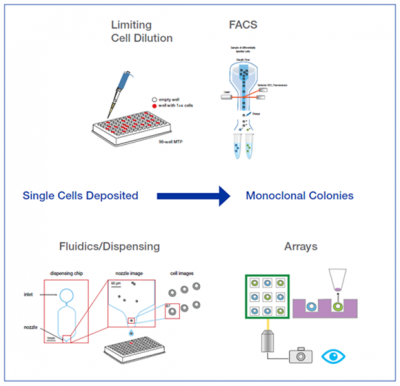
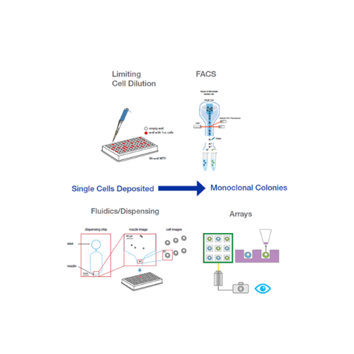
Figure 1: Current methods for single cell cloning. The majority of technologies available on the market separate a heterogeneous population of cells into single cells within a microwell, either via manual pipetting or fluidics-based segregation.
Overcoming Limitations of Current Technologies
CellRaft Technology is an affordable and modern method for developing monoclonal cell lines that overcomes the limitations of traditional methods. It provides flask-like culture conditions at the resolution of a single cell, with gentle and automated isolation using image-based attributes for function, gene expression, and morphology. Within a single platform, researchers can grow, scan, analyze, and isolate single cell derived monoclonal colonies.

Figure 2: CellRaftA microscale polystyrene growth surface within an array used to spatially segregate cells/colonies while maintaining shared media access, enabling imaging over time and targeted isolation. More Technology workflow. The advantage of the CellRaftA microscale polystyrene growth surface within an array used to spatially segregate cells/colonies while maintaining shared media access, enabling imaging over time and targeted isolation. More technology is that cells are seeded as a population while maintaining single-cell separation. Cells can grow in situ on individual CellRafts to form discrete colonies that can be identified using image-based software analysis tools. After growth on the array, the monoclonal colony, rather than a single cell, is isolated intact and transferred to a downstream collection plate for continued growth, expansion, and downstream analysis.
Benefits of CellRaftA microscale polystyrene growth surface within an array used to spatially segregate cells/colonies while maintaining shared media access, enabling imaging over time and targeted isolation. More Technology
The use of CellRaftA microscale polystyrene growth surface within an array used to spatially segregate cells/colonies while maintaining shared media access, enabling imaging over time and targeted isolation. More Technology offers several advantages over traditional methods for developing monoclonal cell lines including:
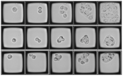
Figure 3: Track-and-trace colony growth from a single cell. Three representative CellRafts containing single cells on day 1 and imaged on the CellRaft AIR SystemAn all-in-one platform for imaging, identifying, and isolating viable single cells, colonies, or organoids using CellRaft Arrays and software-guided selection with CellRaft Cytometry. More over a 4-day period until colony formation. This serial imaging allows for precise monitoring of exponential growth and phenotypic characterization, as well as improved viability downstream.
One of the most critical components of cell line development is proof of monoclonalityEvidence that a colony/line originated from one cell; often supported by time-course imaging and traceability. More, and as the saying goes “a picture is worth a thousand words”. The CellRaftA microscale polystyrene growth surface within an array used to spatially segregate cells/colonies while maintaining shared media access, enabling imaging over time and targeted isolation. More AIR can image an entire CellRaft arrayConsumables containing thousands of microwells (containing CellRafts) for spatial segregation, imaging, and isolation of single cells, monoclonal colonies, or 3D cultures. More in as little as 6 minutes in brightfield, providing a saved image of every single CellRaftA microscale polystyrene growth surface within an array used to spatially segregate cells/colonies while maintaining shared media access, enabling imaging over time and targeted isolation. More for impeccable record keeping and the ability to track and trace single cells from seeding to colony formation (Figure 3). In addition, because the single cells are individually segregated within the array but share a contiguous media volume, viability and clonal growth are significantly improved compared to methods that put a single cell in a well. This key feature leads to hundreds to thousands of clonal colonies to choose from, ensuring that the researchers are able to get exactly the phenotypic characteristics they desire, rather than settling for whichever clones survive. Lastly, the CellRaftA microscale polystyrene growth surface within an array used to spatially segregate cells/colonies while maintaining shared media access, enabling imaging over time and targeted isolation. More technology is user-friendly, easy to learn, and requires a very low barrier to entry compared to instrumentation such as flow sorters.
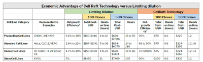
Table 1:
Conclusion
In summary, monoclonal cell line development is an important step in biomedical research that has traditionally relied on time-consuming and labor-intensive techniques or high-cost alternatives that produce low-viability cell populations. However, CellRaftA microscale polystyrene growth surface within an array used to spatially segregate cells/colonies while maintaining shared media access, enabling imaging over time and targeted isolation. More Technology offers significant advantages over traditional methods due to its affordability, speed, accuracy, and scalability capabilities, as well as its overall improvement of cell viability and clonal growth. For scientists struggling to find an efficient and successful method for growing and recovering high-quality monoclonal cell lines, CellRaftA microscale polystyrene growth surface within an array used to spatially segregate cells/colonies while maintaining shared media access, enabling imaging over time and targeted isolation. More Technology is a novel solution that will improve overall research productivity and progress.
For a deeper look comparing technologies, download the white paper, Development of Monoclonal Cell Lines – Available Technologies and Overcoming Challenges.

Kap Kumar has over 25 years of experience in the life sciences tools and reagents industry. He started in R&D and product development, where he launched products for cell biology and imaging applications. For the last 15 years, Kap has led strategic marketing, market development, and product management for a variety of companies, including Thermo Fisher Scientific (Life Technologies), Danaher (Beckman Coulter Life Sciences), Cell Signaling Technologies, Nexcelom Biosciences, and Avantor-VWR. Kap has diverse experience managing complex portfolios, including instruments, consumables, and reagents, both in early-stage and mature companies. Kap has a Ph.D. in Cell and Molecular Biology from Kent State University, a post-doctorate from Harvard Medical School, and an MBA from Babson College.




By Jessica Hartman, Ph.D.
Toiling in the lab
With a deadline on its way
To the scope I go
On a Saturday
I don’t see a clone
I’ve looked for one that’s right
What misery these data bring,
I’ll have to look all night,

Oh! Single cells, single cells, dilutions aren’t the way
Oh, I wish I had an AIR to pick a clone, to-da-ay!
Single cells, single cells, I need a better way
Use a Raft to grow a clone or be sad this holiday!
Use a Raft to grow a clone or be sad this holiday!
Now it’s getting late
My spirits are so low
My PI will hate
If my cells don’t grow
My eyesight’s getting dim
A clone I cannot see
My chances are so slim
I need CellRaftA microscale polystyrene growth surface within an array used to spatially segregate cells/colonies while maintaining shared media access, enabling imaging over time and targeted isolation. More Cytometry
Oh! Single cells, single cells, sorters aren’t the way
Oh, I wish I had an AIR to pick a clone, to-da-ay!
Single cells, single cells, I need a better way
Use a Raft to grow a clone or be sad this holiday!
Use a Raft to grow a clone or be sad this holiday!
Oh! Single cells, single cells, dispensers aren’t the way
Oh, I wish I had an AIR to pick a clone, to-da-ay!
Single cells, single cells, I need a better way
Use a Raft to grow a clone or be sad this holiday!
Use a Raft to grow a clone or be sad this holiday!


“Single Cell” vocalist: Virginia Laurie