
It may be difficult to fathom, but that little device you carry in your pocket to play Candy Crush and read your social media feeds has enough computing power to put supercomputers from just a few decades ago to shame. Similarly, today’s desktop computers have come a long way in raw processing power, artificial intelligence, and machine learning. Now researchers are harnessing this power to improve how we develop critical new drugs faster and cheaper.
In our ultimate goal to find the perfect drugs to cure our ailments, pharma companies have developed massive drug screening facilities to test thousands of potential drugs against the target. The typical screening facility now looks more like an automated manufacturing plant for computer chips.
Despite these advances, the development process is still extremely complex. Although a drug may act perfectly on its intended target, it may have unwanted and unseen consequences somewhere else, known in the industry as toxicity.
A perfect example is cardiotoxicity from off-target drug interactions. Electrical signals in the heart are maintained by proteins in heart cells known as ion channels. A heartbeat is regulated by an intricate interplay as these ion channels conduct sodium, potassium and calcium in and out of the cells. If a drug interferes with this coordinated effort, it can lead to arrhythmias and heart attacks. Several drugs targeting common ailments (e.g. the antihistamine drug terfenadine) were pulled from the market due to safety issues after the discovery of their detrimental interaction with ion channels in the heart (in this case, the hERG ion channel).
To identify these interactions early in the development process, high-throughput assays have been developed to measure drug interactions with hERG and other ion channels. A successful technology used for this is the Automated Patch Clamp (APC), where engineered cells carrying these ion channels are subjected to various drugs and their interactions are quantified.
Read about IonFlux, a microfluidics-based APC and Potassium channels
Although experimental observation is key to understanding hERG-induced cardiotoxicity, computational models using artificial intelligence are now possible. These models have the potential to improve both the speed and efficiency of the current test-based methods. Researchers at the University of Alberta were able to build a computational model that can complement the experimental screening process in successfully predicting effects of drugs based on their molecular structure.
Their ground-breaking research has been described in a Nature Scientific Reports article. The study compares several standard drugs experimentally evaluated with automated patch clamp to their predicted interactions via computational modeling. According to the researchers, “given the results, our workflow would not only be useful for predicting drug-mediated hERG liability but can also be used as a complementary protocol for designing functional groups when optimizing the drug-like leads to reduce their hERG liability.”
A structure-based computational workflow to predict liability and binding modes of small molecules to hERG. |
Dr. Khaled H. Barakat presented the computational model in a webinar shortly after the paper was published. The paper is co-authored with Dr. Michael Houghton, 2020 Nobel Prize Laureate in Physiology or Medicine.
Click to access to prerecorded webinar
Other researchers are attempting to translate this predictive capability from drug development to personalized diagnostics. A recent study uses a combination of experimental results and patient genetic information to adapt the model to each patient. This approach could provide the foundation for a computational model to provide personalized patient treatment for cardiac arrhythmias.
Genetic algorithm-based personalized models of human cardiac action potential D.N. Smirnov et al. PLOS ONE. May.11.2020 |
Applications of multiscale computational models and analytics are not restricted to cardiac disorders. From cancer treatment to brain disease, computational models are providing a better understanding of drug interaction and development. Although still far from perfect, these approaches represent a solid foundation for implementing effective personalized medicine.

Ali Yehia has a Ph.D. in Cardiac Electrophysiology from McGill University. He has been involved in ion channel screening using Automate Patch Clamp since 2006. He joined Fluxion Biosciences in 2012, where he developed ion channel assays and IonFlux Mercury systems. He was the Chief Scientific Officer at Fluxion before moving to Cell Microsystems as the new Senior Director of the IonFlux product.

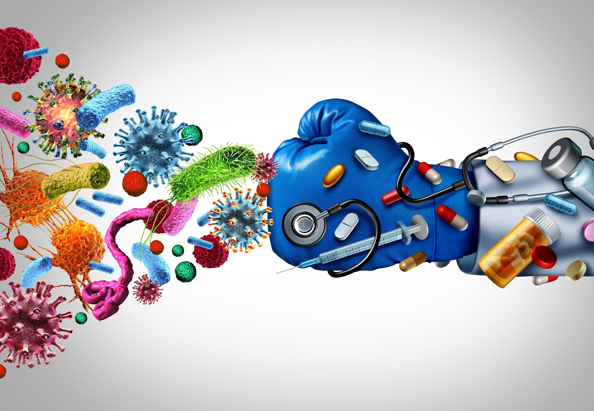
In the ongoing battle against antibiotic-resistant bacteria, or “superbugs,” scientists are constantly seeking innovative tools to understand the mechanisms of resistance and develop effective treatments. One such groundbreaking technology is shear flow systems, which have emerged as game-changers in microbiological research. By simulating the dynamic conditions of bacteria’s natural environments, such as the human body or food processing, shear flow systems provide unique insights into bacterial behavior and resistance mechanisms that traditional static methods simply cannot achieve. In this blog post, we’ll explore the benefits of using shear flow systems to combat antibiotic resistance and develop next-generation therapies.
Antibiotic resistance is a complex phenomenon influenced by a variety of factors, including genetic mutations, horizontal gene transfer, and the formation of biofilms. Traditional laboratory methods often fail to capture the dynamic nature of these processes, leading to incomplete or misleading results. Shear flow systems address this limitation by providing a more realistic environment for bacterial growth and interaction. Here’s how:
1. Mimicking Real-World Conditions
In the human body, bacteria are rarely in a static state. They are constantly exposed to fluid flow, such as blood circulation or urine flow, which influences their behavior and resistance mechanisms. Shear flow systems can replicate these conditions, allowing researchers to study how bacteria respond to antibiotics under realistic physiological conditions. This is critical for understanding how resistance develops and persists in vivo. This benefit was recently highlighted in a webinar presented by Dr. Katharina Richter, a microbiology researcher from the University of Adelaide in Australia. Using a high-throughput BioFlux Shear Flow System, Dr. Richter and her team were able to test 3 different methods of superbug treatments under physiological conditions.
2. Studying Biofilm Formation and Resistance
Biofilms—structured communities of bacteria encased in a protective matrix—are a major contributor to antibiotic resistance. Biofilms are notoriously difficult to treat because they shield bacteria from antibiotics and the immune system. Shear flow systems enable researchers to study biofilm formation in real time, observing how bacteria adhere to surfaces, form microcolonies, and develop resistance under flow conditions. For example, using a BioFlux, Dr. Richter was able to leverage high-resolution imaging to obtain unprecedented insight into the biofilm killing and prevention efficacy of a copper and diethyldithiocarbamate (DDC) combination nanoparticles¹. The group is currently investigating the use of a Cu-DDC infused gel that can be injected into wounds that are at high risk for chronic infection, such as hernia.
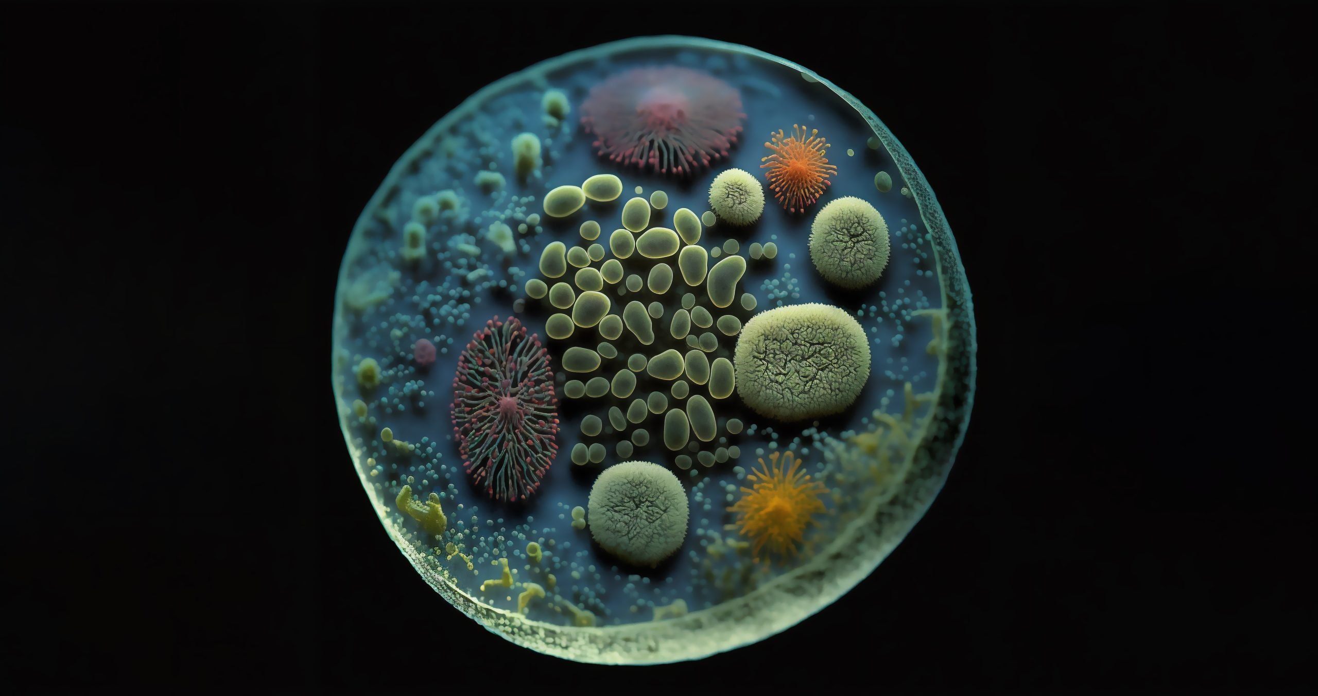
The study of biofilm communities presents unique challenges in experimental design and analysis. While mixed bacterial populations offer fascinating insights into microbial interactions and community dynamics, accurately characterizing and investigating these complex systems has previously required sophisticated and tedious technical approaches. Here, we will discuss advances in technology that have helped to simplify and streamline polymicrobial investigations.
Throughout the last decade, a trend for growing and assaying biofilms under shear flow has gained traction among microbiologists. The rationale for investigating biofilms under flow is multi-dimensional. A recent Cell Microsystems survey asked why microbiologists are using shear flow found that the top responses were:
Of the scientists surveyed, 40% indicated that they use a BioFlux Shear Flow System. This system provides unique advantages over other methods to induce shear stress, such as slide-based parallel plate flow chambers and benchtop rockers.
BioFlux uses microfluidic channels embedded onto the bottom of standard-sized 6, 24, or 48-well plates to create isolated and independent flow chambers where user-defined wall shear stress is initiated and maintained by digitally controlled air pressure. This not only simplifies the setup and experimental processes but also ensures that experiments are contained and do not contaminate the shear flow system.
Although shear flow has been thoroughly demonstrated to improve the biological relevance of biofilm investigations1, an often overlooked aspect of biofilm physiology is the significant role that multicellular aggregates play in biofilm formation2. When investigating Pseudomonas aeruginosa, it has been demonstrated that multicellular aggregates lead to faster biofilm formation and enhanced resistance to antimicrobials and immune responses3. In addition, multispecies microbial aggregates have been shown to dominate single cells in human saliva4. Together, these data show that aggregates are important areas of investigation because they are more likely to represent physiological biofilm formation and growth.