Bacterial biofilms are found throughout nature, in places such as ponds, the human body, and food. Although humans peacefully co-exist and even benefit from many types of bacteria, some strains, such as Pseudomonas aeruginosa, can form pathogenic biofilms. To combat these harmful biofilms, many researchers are engaged in investigations aimed at determining the mechanisms of antimicrobial resistance and/or discovering and developing antimicrobial materials and pharmaceuticals.
Often, the translation of the results of antimicrobial investigations to real-world scenarios is disappointing, with many materials and pharmaceuticals not living up to expectations that were set during lab experimentation. A major factor in these real-world failures is that many biofilms are investigated under static conditions, which do not represent how biofilms exist in real-world environments. Here we highlight recent, high-impact, peer-reviewed articles from scientists who improved the physiological relevance of their antimicrobial investigations by assaying biofilms under shear stress.
In a fascinating investigation, Rostami et al. delved into the hidden battleground within our mouths where bacteria engage in fierce competition for survival. The study focused on a specific bacterium called Streptococcus gordonii and its weapon of choice: the enzyme SsnA. By growing mixed-species biofilms under low shear flow of primary saliva, the group found that SsnA works by breaking down the DNA released by other bacteria, disrupting their ability to form biofilms and survive in the oral environment (Figure 1). This discovery sheds light on the complex interactions between different species of bacteria in our mouths and how they compete for resources and territory. Understanding these dynamics could lead to new strategies for promoting oral health and combating diseases like tooth decay and gum inflammation.
Journal: NPJ Biofilms and Microbiomes, December 2022
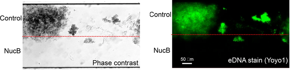
Figure 1.Sucrose (0.3%) supplemented saliva was used to grow microcosm biofilms and Bacillus licheniformis extracellular nuclease NucB was included in the lower half of the growth channel. Adapted from Rostami et al. 2022. doi.org/10.1038/s41522- 022-00359-z
In an exciting study by Kaul et al., consistent feeding and waste removal provided by shear flow enabled the group to grow truly mature biofilms, empowering the discovery of a powerful weapon in the fight against stubborn bacterial infections (Figure 2). Although mature biofilms formed by Staphylococcus aureus and Staphylococcus epidermidis are notorious for being tough and antibiotic-resistant, the researchers found that by combining two substances – diethyldithiocarbamate (DETC) and copper ions – they effectively destroyed these bacterial biofilms. When used together, DETC and copper ions acted like a dynamic duo, penetrating the biofilms and killing the bacteria within. This combination was effective both in laboratory tests and in live animal models, showing promise for future treatments of bacterial infections.
Journal: Frontiers in Microbiology, September 2022
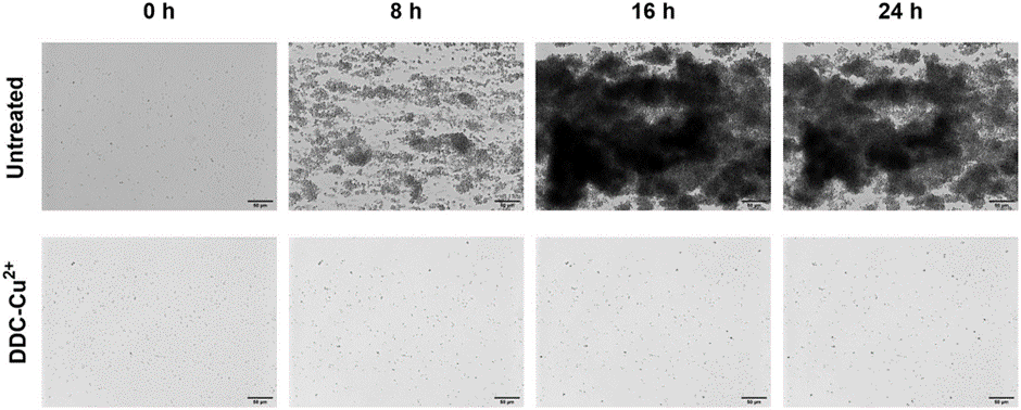
Figure 2. Monitoring of MRSA Mu50 biofilm formation over 24 h when left untreated or treated with a combination of 8 μg/mL diethyldithiocarbamate and 32 μg/mL Cu2+ combination (DDC-Cu2+) using the Bioflux system. Scale bar represents 50 μm. Kaul et al. 2022. doi.org/10.3389/fmicb.2022.999893
In an important investigation, Pouget et al. made strides in the battle against infections in diabetic foot wounds. The study focused on two troublesome bacteria commonly found in diabetic foot wounds: Staphylococcus aureus and Pseudomonas aeruginosa, which are known to create antibiotic-resistant biofilms. The group found that traditional antibiotics, while effective against these planktonic bacteria in a liquid environment, struggle to penetrate and kill the bacteria within biofilms. To address this challenge, the researchers developed a novel microfluidic model that mimics the environment of a diabetic foot wound. Using this model, they tested the antimicrobial activity of different antibiotics against biofilms formed by S. aureus and P. aeruginosa. Their findings revealed that while some antibiotics were able to partially inhibit biofilm formation, none were able to completely eradicate the biofilms. This underscores the need for new strategies to combat bacterial biofilms in diabetic foot wounds.
Journal: Journal of Antimicrobial Chemotherapy, February 2023
In a recent study, de Jongh et al. uncovered a surprising collaboration between two common microorganisms found in the mouth: Porphyromonas gingivalis, a type of bacteria, and Candida albicans, a type of fungus. Both are known to colonize the mouth and contribute to oral infections, but their interactions are not fully understood. Leveraging a microfluidic shear flow system, the group mimicked the saliva flow of the oral cavity to co-culture P. gingivalis and C. albicans (Figure 3). The researchers discovered that when these two microorganisms come into contact, they form a symbiotic relationship that enhances their ability to adhere to surfaces in the mouth and cause harm. One of the study’s key findings was that the presence of C. albicans allows P. gingivalis to thrive in oxygen-rich environments, which are normally toxic to the bacteria. This enables P. gingivalis to spread more easily in the mouth, contributing to the progression of gum disease. Their partnership, while unexpected, has profound implications for oral health and may contribute to conditions like gum disease. Overall, this research provides valuable insights into the complex interactions between microorganisms in the mouth and highlights the importance of understanding these dynamics for maintaining oral health.
Journal: Biofilms, June 2024
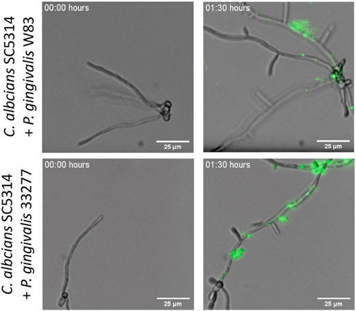
Figure 3. Adherence of P. gingivalis labeled with carboxyfluorescein succinimidyl ester (CFSE) to C. albicans. P. gingivalis adheres to C. albicans hyphae within 1.5 h. Scale bar indicates 25 μm in each picture. Adapted from de Jongh et al. 2024. doi.org/10.1016/j. bioflm.2023.100172
5. Anti-biofilm activity of murepavadin against cystic fibrosis Pseudomonas aeruginosa isolates
Recently, Díez-Aguilar et al. made what may be a significant breakthrough in the fight against infections caused by Pseudomonas aeruginosa in individuals with cystic fibrosis (CF). P. aeruginosa is a common and stubborn bacterium that can cause serious lung infections in people with CF, leading to respiratory problems and other health complications. The group examined the impact of 4 antibiotics on 53 morphotypes under both static and shear flow conditions. Although all of the tested antibiotics were effective at high concentrations, interestingly, the concentration of aztreonam required for antibiofilm activity was much lower when used under shear flow compared to static cultures. However, the researchers found that a compound called murepavadin showed the most promising anti-biofilm activity against P. aeruginosa isolates. Murepavadin penetrated the biofilms and effectively killed the bacteria, even those that were resistant to traditional antibiotics. This discovery offers hope for new treatment options for individuals with CF who are battling P. aeruginosa infections. By targeting biofilms with compounds like murepavadin, scientists may overcome the challenges posed by antibiotic resistance and improve patient outcomes.
Journal: Journal of Antimicrobial Chemotherapy, September 2021
Conclusion
Culturing and assaying bacterial biofilms under shear flow conditions not only induces changes in response to shear stress but also improves feeding and waste removal. Together, these factors enable the growth of truly mature biofilms that better represent biofilms in their natural environments. Using the totally complete, easy-to-use, cost-effective BioFlux shear flow system enabled these investigators with the ability to enhance the physiological relevance of their antimicrobial investigations.

Dr. Anson Blanks completed his BS in exercise physiology at East Carolina University and his MS in clinical exercise science at Appalachian State University. After working as a clinical exercise physiologist in cardiopulmonary rehabilitation, Dr. Blanks decided to pursue a career in scientific research. He attended Virginia Commonwealth University, where he completed his Ph.D. in Rehabilitation and Movement Science. After spending several years as a research and development scientist in biotechnology industry, Dr. Blanks is now a scientific marketing manager for Cell Microsystems in Durham, NC.


In the ongoing battle against antibiotic-resistant bacteria, or “superbugs,” scientists are constantly seeking innovative tools to understand the mechanisms of resistance and develop effective treatments. One such groundbreaking technology is shear flow systems, which have emerged as game-changers in microbiological research. By simulating the dynamic conditions of bacteria’s natural environments, such as the human body or food processing, shear flow systems provide unique insights into bacterial behavior and resistance mechanisms that traditional static methods simply cannot achieve. In this blog post, we’ll explore the benefits of using shear flow systems to combat antibiotic resistance and develop next-generation therapies.
Antibiotic resistance is a complex phenomenon influenced by a variety of factors, including genetic mutations, horizontal gene transfer, and the formation of biofilms. Traditional laboratory methods often fail to capture the dynamic nature of these processes, leading to incomplete or misleading results. Shear flow systems address this limitation by providing a more realistic environment for bacterial growth and interaction. Here’s how:
1. Mimicking Real-World Conditions
In the human body, bacteria are rarely in a static state. They are constantly exposed to fluid flow, such as blood circulation or urine flow, which influences their behavior and resistance mechanisms. Shear flow systems can replicate these conditions, allowing researchers to study how bacteria respond to antibiotics under realistic physiological conditions. This is critical for understanding how resistance develops and persists in vivo. This benefit was recently highlighted in a webinar presented by Dr. Katharina Richter, a microbiology researcher from the University of Adelaide in Australia. Using a high-throughput BioFlux Shear Flow System, Dr. Richter and her team were able to test 3 different methods of superbug treatments under physiological conditions.
2. Studying Biofilm Formation and Resistance
Biofilms—structured communities of bacteria encased in a protective matrix—are a major contributor to antibiotic resistance. Biofilms are notoriously difficult to treat because they shield bacteria from antibiotics and the immune system. Shear flow systems enable researchers to study biofilm formation in real time, observing how bacteria adhere to surfaces, form microcolonies, and develop resistance under flow conditions. For example, using a BioFlux, Dr. Richter was able to leverage high-resolution imaging to obtain unprecedented insight into the biofilm killing and prevention efficacy of a copper and diethyldithiocarbamate (DDC) combination nanoparticles¹. The group is currently investigating the use of a Cu-DDC infused gel that can be injected into wounds that are at high risk for chronic infection, such as hernia.
The study of biofilm communities presents unique challenges in experimental design and analysis. While mixed bacterial populations offer fascinating insights into microbial interactions and community dynamics, accurately characterizing and investigating these complex systems has previously required sophisticated and tedious technical approaches. Here, we will discuss advances in technology that have helped to simplify and streamline polymicrobial investigations.
Throughout the last decade, a trend for growing and assaying biofilms under shear flow has gained traction among microbiologists. The rationale for investigating biofilms under flow is multi-dimensional. A recent Cell Microsystems survey asked why microbiologists are using shear flow found that the top responses were:
Of the scientists surveyed, 40% indicated that they use a BioFlux Shear Flow System. This system provides unique advantages over other methods to induce shear stress, such as slide-based parallel plate flow chambers and benchtop rockers.
BioFlux uses microfluidic channels embedded onto the bottom of standard-sized 6, 24, or 48-well plates to create isolated and independent flow chambers where user-defined wall shear stress is initiated and maintained by digitally controlled air pressure. This not only simplifies the setup and experimental processes but also ensures that experiments are contained and do not contaminate the shear flow system.
Although shear flow has been thoroughly demonstrated to improve the biological relevance of biofilm investigations1, an often overlooked aspect of biofilm physiology is the significant role that multicellular aggregates play in biofilm formation2. When investigating Pseudomonas aeruginosa, it has been demonstrated that multicellular aggregates lead to faster biofilm formation and enhanced resistance to antimicrobials and immune responses3. In addition, multispecies microbial aggregates have been shown to dominate single cells in human saliva4. Together, these data show that aggregates are important areas of investigation because they are more likely to represent physiological biofilm formation and growth.