


Recently, some members of the Cell Microsystems team traveled to the United Kingdom, to speak with researchers at some of the institutions associated with the National Biofilms Innovation Center (NBIC). While there, our team was fortunate to speak with several microbiologists who were conducting investigations in two major areas: biofilm drug resistance and antibiofilm drug development. In this blog post, we highlight these investigators’ approach to overcoming common obstacles in biofilm research.
Drug Resistance & Development of Antibiofilm Treatments
A major reason that microorganisms, such as bacteria and fungi, form biofilms is to protect themselves from external threats. This often means that complete penetration of biofilms by antibiofilm treatments is difficult, particularly in areas such as the lungs or inside of cells. To overcome the protective effects of biofilms, researchers at institutions associated with the NBIC are examining new methods of antibiofilm drug delivery. One of the major drug delivery methods currently under investigation is the use of nanoparticles. As the name suggests, nanoparticles are very small in size, typically ranging from 1-100 nm. Although certain types of nanoparticles exist in nature, investigators at NBIC institutions have crafted synthetic nanoparticles with antibiofilm drugs inside of them. The advantage of this method is that biofilm-harboring cells take up the nanoparticles and then the antibiofilm drug is released, delivering treatment directly to microbes within the biofilm. This method prevents beneficial microorganisms from being exposed to potentially harmful treatment. The investigators at NBIC institutions hope this targeted approach can reduce the amount of drug required to effectively treat intracellular biofilms and preserve a healthy microbiome.
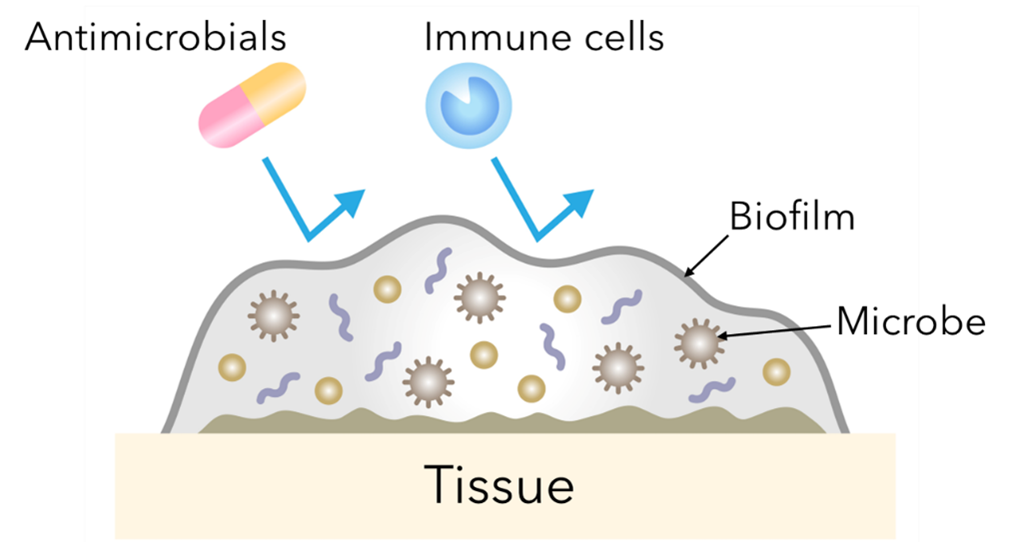
Figure 1. The protective effects of biofilms. This figure highlights how microbes form biofilms to help protect themselves from external threats, such as immune cells and antimicrobial treatments.

Figure 2. Mechanisms of antibiotic resistance. Known mechanisms of how bacteria can reduce or eliminate their responsiveness to antibiotic treatments.
Challenges in Biofilm Investigation
Traditionally, to grow microbial strains of interest, microbiologists follow the basic steps outlined below:
This workflow requires a significant time investment, causes changes in biofilm morphology due to compression by the coverslip, has an imaging throughput of a single biofilm, and perhaps most frustratingly, often yields results that do not transfer to in vivo models.
Most biofilms, particularly those that form within the body, grow under some form of liquid flow. Therefore, shear flow chambers represent an improvement over the traditional workflow by enabling the analysis of biofilms in biological conditions. This can create major benefits, such as improved nutrition for the entire biofilm2 and enhanced biofilm attachment3. Therefore, to overcome the obstacle of in vitro to in vivo translation, some microbiology labs have developed their own shear flow chambers to conduct biofilm assays.
Creating a DIY setup, such as a parallel plate flow chamber, typically involves purchasing a peristaltic pump, creating a specialized flow chamber, attaching a liquid reservoir, creating a pressure sensor, attaching resistance valves, attaching tubing, and running the setup through a dampener. Although these components can be purchased separately, assembly, calibration, cleaning, and maintenance are difficult and time-consuming. In addition, control of the flow rate is difficult and imaging under flow is extremely challenging. Furthermore, the throughput of DIY shear flow systems is often limited to 1-6 samples. This can be a dealbreaker for many labs, as a recent survey conducted by Cell Microsystems found that 60% of respondents required between 11 and 49 samples for their projects to be successful.
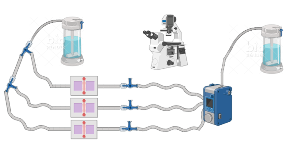
Figure 3. Simplified “DIY” flow cell setup. A typical setup includes flow chamber(s), reservoirs, tubing, 3-way stopcocks, male and female luer adapters, a pressure controller, and a microscope with a camera for imaging.
Despite the benefits of assaying biofilms under shear flow, shear flow devices have not been widely adopted by microbiology labs. In a recent CMS survey of microbiologists, it was found that 100% of respondents were aware that shear flow provides benefits to biofilm investigations; however, only 40% currently use some form of shear flow in their investigations. This is likely due to the difficulty of creating and maintaining a “do-it-yourself” shear flow chamber.
A Shear Flow Solution
To enhance the biological relevance of their biofilm investigations, while also achieving the throughput they need, investigators at many of the institutions associated with the NBIC have adopted BioFlux shear flow systems. The BioFlux systems enable the culture, assay, and imaging of biofilms under shear flow conditions.
BioFlux presents several improvements over DIY flow chambers, including being a complete system with a simple setup that requires no calibration and is easy to clean because the liquid flow is controlled by pneumatic air pressure, not liquid flow through tubes. The shear flow rate is also easily calculated and controlled with a click in the BioFlux software. Additionally, BioFlux eliminates “fluid bursts” that occur at the startup of peristaltic pumps, which can lead to undesired biofilm detachment. Perhaps the most beneficial feature of BioFlux are the microfluidic plates. BioFlux plates are Society for Biomolecular Screening (SBS) standard-sized plates with microfluidic channels embedded into the bottom of the plates, with coverslip glass viewing windows. This means that investigators can leverage up to 24 sequential experiment throughput using most inverted microscopes that are compatible with standard-sized microplates. BioFlux has enabled high-resolution imaging and video creation of non-deformed biofilms under shear flow by investigators at these NBIC affiliated institutions.
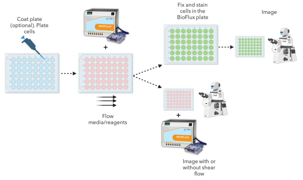
Figure 4. Biofilm workflow using BioFlux. A general workflow showing how biofilms can be grown and assayed using a BioFlux system. Biofilms can be fixed and imaged or imaged live while under shear flow.
The microbiologists that we interviewed stressed how the experimental throughput combined with the standardization and reproducibility of BioFlux has greatly improved the workflow of their labs. Using this microfluidic shear flow technology, these investigators are able to test a similar number of biofilms as a static workflow, and much more than a DIY shear flow system, while leveraging the benefits of a biologically relevant environment. Together, these benefits have and will continue to empower microbiology labs to accelerate biofilm and antimicrobial discoveries.
Enhance your biofilm investigations
References

Dr. Anson Blanks completed his BS in exercise physiology at East Carolina University and his MS in clinical exercise science at Appalachian State University. After working as a clinical exercise physiologist in cardiopulmonary rehabilitation, Dr. Blanks decided to pursue a career in scientific research. He attended Virginia Commonwealth University, where he completed his Ph.D. in Rehabilitation and Movement Science. After spending several years as a research and development scientist in biotechnology industry, Dr. Blanks is now a scientific marketing manager for Cell Microsystems in Durham, NC.


In the ongoing battle against antibiotic-resistant bacteria, or “superbugs,” scientists are constantly seeking innovative tools to understand the mechanisms of resistance and develop effective treatments. One such groundbreaking technology is shear flow systems, which have emerged as game-changers in microbiological research. By simulating the dynamic conditions of bacteria’s natural environments, such as the human body or food processing, shear flow systems provide unique insights into bacterial behavior and resistance mechanisms that traditional static methods simply cannot achieve. In this blog post, we’ll explore the benefits of using shear flow systems to combat antibiotic resistance and develop next-generation therapies.
Antibiotic resistance is a complex phenomenon influenced by a variety of factors, including genetic mutations, horizontal gene transfer, and the formation of biofilms. Traditional laboratory methods often fail to capture the dynamic nature of these processes, leading to incomplete or misleading results. Shear flow systems address this limitation by providing a more realistic environment for bacterial growth and interaction. Here’s how:
1. Mimicking Real-World Conditions
In the human body, bacteria are rarely in a static state. They are constantly exposed to fluid flow, such as blood circulation or urine flow, which influences their behavior and resistance mechanisms. Shear flow systems can replicate these conditions, allowing researchers to study how bacteria respond to antibiotics under realistic physiological conditions. This is critical for understanding how resistance develops and persists in vivo. This benefit was recently highlighted in a webinar presented by Dr. Katharina Richter, a microbiology researcher from the University of Adelaide in Australia. Using a high-throughput BioFlux Shear Flow System, Dr. Richter and her team were able to test 3 different methods of superbug treatments under physiological conditions.
2. Studying Biofilm Formation and Resistance
Biofilms—structured communities of bacteria encased in a protective matrix—are a major contributor to antibiotic resistance. Biofilms are notoriously difficult to treat because they shield bacteria from antibiotics and the immune system. Shear flow systems enable researchers to study biofilm formation in real time, observing how bacteria adhere to surfaces, form microcolonies, and develop resistance under flow conditions. For example, using a BioFlux, Dr. Richter was able to leverage high-resolution imaging to obtain unprecedented insight into the biofilm killing and prevention efficacy of a copper and diethyldithiocarbamate (DDC) combination nanoparticles¹. The group is currently investigating the use of a Cu-DDC infused gel that can be injected into wounds that are at high risk for chronic infection, such as hernia.
The study of biofilm communities presents unique challenges in experimental design and analysis. While mixed bacterial populations offer fascinating insights into microbial interactions and community dynamics, accurately characterizing and investigating these complex systems has previously required sophisticated and tedious technical approaches. Here, we will discuss advances in technology that have helped to simplify and streamline polymicrobial investigations.
Throughout the last decade, a trend for growing and assaying biofilms under shear flow has gained traction among microbiologists. The rationale for investigating biofilms under flow is multi-dimensional. A recent Cell Microsystems survey asked why microbiologists are using shear flow found that the top responses were:
Of the scientists surveyed, 40% indicated that they use a BioFlux Shear Flow System. This system provides unique advantages over other methods to induce shear stress, such as slide-based parallel plate flow chambers and benchtop rockers.
BioFlux uses microfluidic channels embedded onto the bottom of standard-sized 6, 24, or 48-well plates to create isolated and independent flow chambers where user-defined wall shear stress is initiated and maintained by digitally controlled air pressure. This not only simplifies the setup and experimental processes but also ensures that experiments are contained and do not contaminate the shear flow system.
Although shear flow has been thoroughly demonstrated to improve the biological relevance of biofilm investigations1, an often overlooked aspect of biofilm physiology is the significant role that multicellular aggregates play in biofilm formation2. When investigating Pseudomonas aeruginosa, it has been demonstrated that multicellular aggregates lead to faster biofilm formation and enhanced resistance to antimicrobials and immune responses3. In addition, multispecies microbial aggregates have been shown to dominate single cells in human saliva4. Together, these data show that aggregates are important areas of investigation because they are more likely to represent physiological biofilm formation and growth.