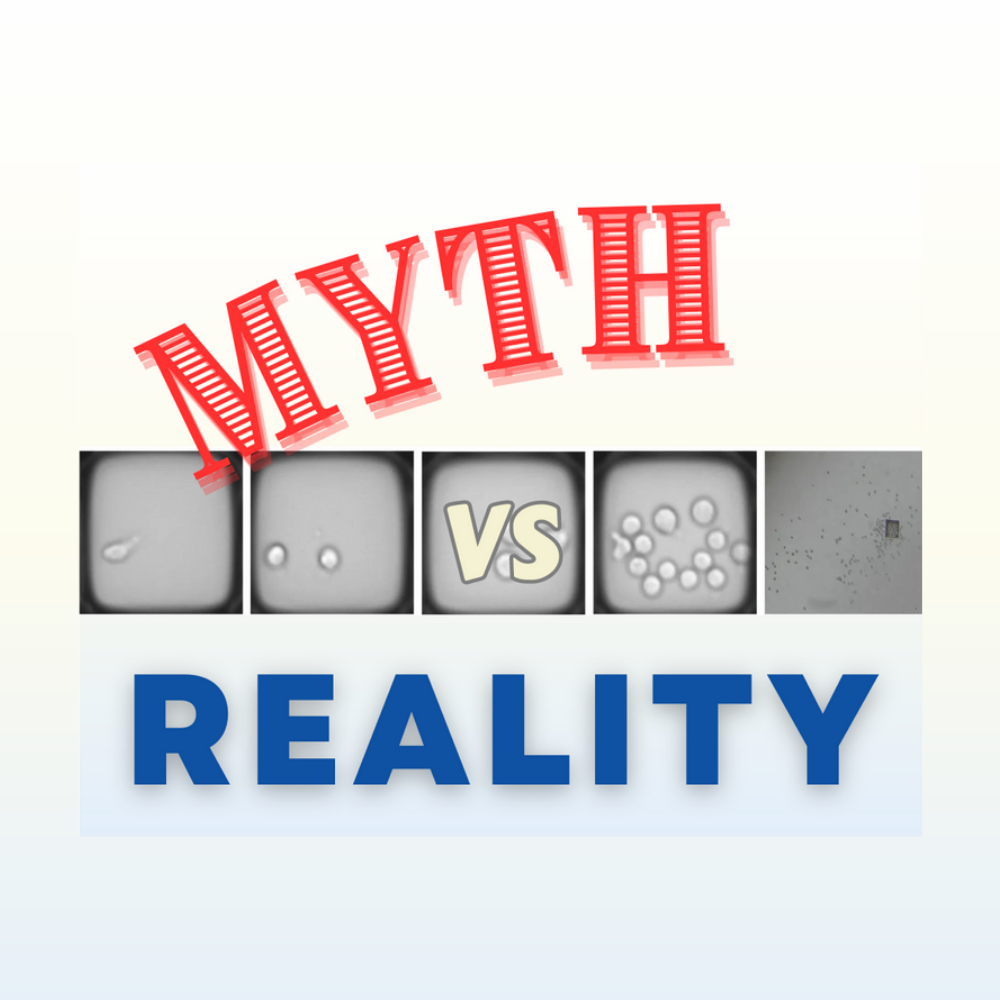
Generating monoclonal colonies is a fundamental step in cell biology research, allowing researchers to study individual cells and their unique characteristics. However, misconceptions and challenges have surrounded this process for years. Fortunately, CellRaft® Technology has emerged as a revolutionary solution, debunking these myths and transforming the way monoclonal colonies are generated. In this blog post, we will explore the top 10 myths associated with generating monoclonal colonies and how CellRaftA microscale polystyrene growth surface within an array used to spatially segregate cells/colonies while maintaining shared media access, enabling imaging over time and targeted isolation. More Technology shatters these barriers, paving the way for more efficient and reliable research.
CellRaftA microscale polystyrene growth surface within an array used to spatially segregate cells/colonies while maintaining shared media access, enabling imaging over time and targeted isolation. More Technology streamlines the process of generating monoclonal colonies by enabling high-throughput single-cell isolation and culture, reducing time and labor required.
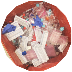
CellRaftA microscale polystyrene growth surface within an array used to spatially segregate cells/colonies while maintaining shared media access, enabling imaging over time and targeted isolation. More Technology optimizes resource utilization by isolating and culturing thousands of individual cells on arrays made up of thousands of microwells that allow cells to be spatially separated while sharing media, minimizing the need for excessive media and plasticware.
CellRaftA microscale polystyrene growth surface within an array used to spatially segregate cells/colonies while maintaining shared media access, enabling imaging over time and targeted isolation. More Technology ensures high cloning efficiency by providing a gentle and non-invasive method of isolating single cells, minimizing stress, and preserving cell viability.

CellRaftA microscale polystyrene growth surface within an array used to spatially segregate cells/colonies while maintaining shared media access, enabling imaging over time and targeted isolation. More Technology enables the imaging of cells from the single cell stage to the colony stage to provide a record of growth.
CellRaftA microscale polystyrene growth surface within an array used to spatially segregate cells/colonies while maintaining shared media access, enabling imaging over time and targeted isolation. More Technology offers a standardized and automated approach, reducing subjective interpretation and minimizing human error.
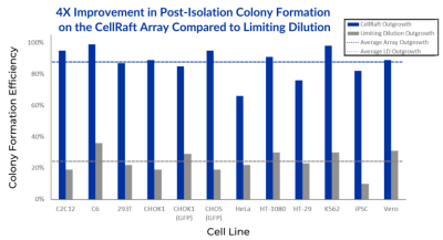
CellRaftA microscale polystyrene growth surface within an array used to spatially segregate cells/colonies while maintaining shared media access, enabling imaging over time and targeted isolation. More Technology preserves high cell viability during subcloning by providing a gentle and controlled environment for isolation, without using fluidics.
CellRaftA microscale polystyrene growth surface within an array used to spatially segregate cells/colonies while maintaining shared media access, enabling imaging over time and targeted isolation. More Technology incorporates traceability features, allowing researchers to easily track and document the lineage of monoclonal colonies, ensuring reproducibility and data integrity.
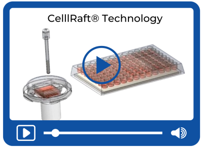
CellRaftA microscale polystyrene growth surface within an array used to spatially segregate cells/colonies while maintaining shared media access, enabling imaging over time and targeted isolation. More Technology enables high-throughput single-cell isolation and culture, facilitating the generation of large numbers of monoclonal colonies with scalability and efficiency.
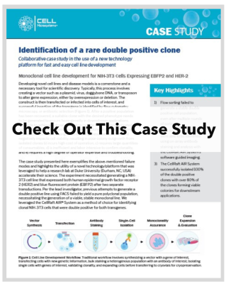
CellRaftA microscale polystyrene growth surface within an array used to spatially segregate cells/colonies while maintaining shared media access, enabling imaging over time and targeted isolation. More Technology excels in identifying and isolating rare cell populations, enabling their characterization and downstream analysis with ease.
CellRaftA microscale polystyrene growth surface within an array used to spatially segregate cells/colonies while maintaining shared media access, enabling imaging over time and targeted isolation. More Technology simplifies the process of generating monoclonal colonies, making it accessible to researchers with varying levels of expertise and reducing the need for specialized skills.
CellRaftA microscale polystyrene growth surface within an array used to spatially segregate cells/colonies while maintaining shared media access, enabling imaging over time and targeted isolation. More Technology has revolutionized the field of generating monoclonal colonies by dispelling the myths and limitations that have hindered this process for years. By offering high-throughput single-cell isolation, optimizing resource utilization, and simplifying workflows, CellRaftA microscale polystyrene growth surface within an array used to spatially segregate cells/colonies while maintaining shared media access, enabling imaging over time and targeted isolation. More Technology empowers researchers to generate monoclonal colonies with enhanced efficiency, reproducibility, and scalability. Embrace the possibilities that CellRaftA microscale polystyrene growth surface within an array used to spatially segregate cells/colonies while maintaining shared media access, enabling imaging over time and targeted isolation. More Technology brings, and unlock new avenues of discovery in cell biology research. Say goodbye to myths and welcome a new era of reliable and impactful research.


Kap Kumar has over 25 years of experience in the life sciences tools and reagents industry. He started in R&D and product development, where he launched products for cell biology and imaging applications. For the last 15 years, Kap has led strategic marketing, market development, and product management for a variety of companies, including Thermo Fisher Scientific (Life Technologies), Danaher (Beckman Coulter Life Sciences), Cell Signaling Technologies, Nexcelom Biosciences, and Avantor-VWR. Kap has diverse experience managing complex portfolios, including instruments, consumables, and reagents, both in early-stage and mature companies. Kap has a Ph.D. in Cell and Molecular Biology from Kent State University, a post-doctorate from Harvard Medical School, and an MBA from Babson College.