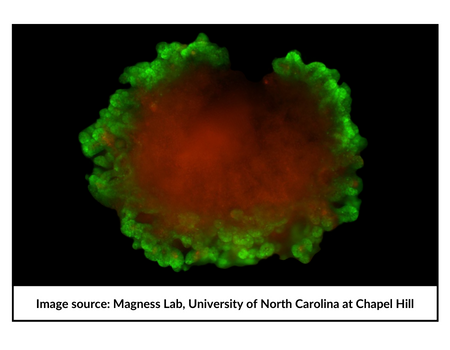
IPSCs and organoids offer a powerful platform for modeling disease & drug discovery. These models can deliver accurate insights which cannot be provided by immortalized cell lines or primary cells and are not subject to limitations of animal models. Notable disease models have been developed for cystic fibrosis, Alzheimer’s disease, and cancer.
The focus of this blog is a key lab and users of the CellRaftA microscale polystyrene growth surface within an array used to spatially segregate cells/colonies while maintaining shared media access, enabling imaging over time and targeted isolation. More technology who use the CellRaftA microscale polystyrene growth surface within an array used to spatially segregate cells/colonies while maintaining shared media access, enabling imaging over time and targeted isolation. More AIR platform as a unique tool for development of novel disease models. The primary focus of this research group centers around the genetics and molecular biology of the GI stem cell. Their aim is to investigate the genes that regulate intestinal stem cell maintenance and differentiation and how these mechanisms impact renewal of the intestinal epithelium in physiology and disease. This information is then used to develop new methods, innovative tools and high throughput platforms to model the human gut ex-vivo. Dr. Scott Magness and his team have developed the art and science of iPSC engineering and organoid generation and utilize the unique features of Cell Raft AIR system as a workhorse platform.
Below is the excerpt of a brief interview with Dr. Magness:
Q: How long have you been using the CellRaft AIR systemAn all-in-one platform for imaging, identifying, and isolating viable single cells, colonies, or organoids using CellRaft Arrays and software-guided selection with CellRaft Cytometry. More and technology?
A: Two years
Q: Why is this platform a game-changer?
A: Prior to the CellRaft Air SystemAn all-in-one platform for imaging, identifying, and isolating viable single cells, colonies, or organoids using CellRaft Arrays and software-guided selection with CellRaft Cytometry. More we were manually identifying single organoids and isolating them. Because of the long times required for these two processes, it was not an option for isolating more than 10 or more organoids. We can now easily identify thousands of single organoids with various morphologies (size, shape, complexity) and isolate hundreds in less than an hour. We also use the CellRaft AIR SystemAn all-in-one platform for imaging, identifying, and isolating viable single cells, colonies, or organoids using CellRaft Arrays and software-guided selection with CellRaft Cytometry. More for cloning of transgenic or CRISPR-edited human intestinal stem cells. Prior to the air system, the efficiency of isolating clonal lines was very inefficient because of the low throughput. Now we can isolate hundreds or thousands of clonal organoids in an unprecedented short amount of time compared to before.
Q: What were you using before the Cell Raft AIR?
A: We were manually isolating either from conventional dome cultures or from the CellRaftA microscale polystyrene growth surface within an array used to spatially segregate cells/colonies while maintaining shared media access, enabling imaging over time and targeted isolation. More arrays before the CellRaft AIR SystemAn all-in-one platform for imaging, identifying, and isolating viable single cells, colonies, or organoids using CellRaft Arrays and software-guided selection with CellRaft Cytometry. More was installed in my lab.
Q: Why would you recommend it to the others working on Organoids or Stem cell engineering?
A: Because organoid heterogeneity is high, this allows us to evaluate many different organoids individually to understand the distribution of heterogeneity in an organoid population. We do this using morphology (site, shape, complexity, or fluorescent reporter genes) or gene expression by qPCR or RNAseq. Additionally, the AIR system is highly efficient for cloning transgenic or gene-edited clones.
Q: What is the unique benefit of the platform that is not seen in other instruments or technologies?
A: The CellRaft ArrayConsumables containing thousands of microwells (containing CellRafts) for spatial segregation, imaging, and isolation of single cells, monoclonal colonies, or 3D cultures. More puts thousands of stem cells or organoids on the same z-plane which drastically improves the imaging and image analysis. The small footprint of the CellRaft ArrayConsumables containing thousands of microwells (containing CellRafts) for spatial segregation, imaging, and isolation of single cells, monoclonal colonies, or 3D cultures. More for culturing drastically reduces costs of reagents such as media and expensive growth factors. The fast imaging of the array on the CellRaft AIR systemAn all-in-one platform for imaging, identifying, and isolating viable single cells, colonies, or organoids using CellRaft Arrays and software-guided selection with CellRaft Cytometry. More enables near real-time imaging of how cells are behaving in response to experimental perturbations. The CellRaftA microscale polystyrene growth surface within an array used to spatially segregate cells/colonies while maintaining shared media access, enabling imaging over time and targeted isolation. More Cytometry feature identifies cells on each CellRaftA microscale polystyrene growth surface within an array used to spatially segregate cells/colonies while maintaining shared media access, enabling imaging over time and targeted isolation. More in the array. These features combined with the ability to automatically isolate cells and organoids into larger conventional well plates (96-well plates) create an integrated workflow that I have not seen in any other system.
Q: How do you see the future of Stem Cell and 3D Biology being impacted due to the CellRaft AIR SystemAn all-in-one platform for imaging, identifying, and isolating viable single cells, colonies, or organoids using CellRaft Arrays and software-guided selection with CellRaft Cytometry. More?
A: We are currently developing high throughput methods on the CellRaft AIR SystemAn all-in-one platform for imaging, identifying, and isolating viable single cells, colonies, or organoids using CellRaft Arrays and software-guided selection with CellRaft Cytometry. More to perform single organoids RNAseq on clonal organoids derived from tumor cells. This is an important technical advance to investigate tumor cell heterogeneity and evolution.
See the latest news from the Magness Lab.
Learn more about CellRaft Technology.

Scott T. Magness, Ph.D.
Associate Professor
University of North Carolina – Chapel Hill,
NC State University Joint Departments of Biomedical Engineering
UNC Departments of Medicine, Cell Biology & Physiology
About the lead Investigator: Dr. Magness is an Associate Professor in the UNC/NCSU joint Departments of Biomedical Engineering. His research is focused on investigating the genetic mechanisms that control the behavior of intestinal and colonic stem cells in health, injury and disease states including inflammatory bowel disease and cancer. Dr. Magness’ group pioneered new intestinal “organoid” technologies in the USA, and has developed a number of platforms that utilize stem cells derived from organ transplant donors or patient biopsies for basic/translational science and commercial applications. For this work, Dr. Magness was given a Transformative Research Award from the National Institutes of Health to develop a human intestinal simulacrum (or mimic) with UNC collaborators. The technology derived from this research was spun-out into Altis Biosystems Inc., which is a hybrid biological tools/CRO focused on providing the pharmaceutical industry with better preclinical cell culture models of the human gut to test drug absorption, secretion, and toxicology.